


Overview
Arthritis is inflammation in or around a joint, where two bones meet. A healthy joint is cushioned by cartilage and lined with synovium tissue which produces fluid to reduce friction during movement. Osteoarthritis occurs when wear and tear of the joint causes breakdown of the cartilage and inflammation of the synovial fluid. Osteoarthritis results from long term use of the joint and is associated with old age. Rheumatoid arthritis also results in breakdown of the joint, but as a result of an auto-inflammatory response.
Current methods to assess arthritis in animal models typically involve external measurements of limbs such as paw volume, paw thickness, and redness. While these methods do determine the overall progression of disease, they do not examine the underlying biology associated with the visible effects of the disease. Since arthritis involves not only inflammation pathways but also changes in vasculature and bone morphology, using fluorescent probes can measure each of these changes individually allow for a more complete assessment of the overall pathology. Preclinical imaging in the far-infrared region allows for non-invasive assessment of the biological response of the mouse over realistic timeframes of disease progression and for monitoring therapeutic efficacy.
Products for imaging arthritis
Our preclinical IVISense™ fluorescent imaging probes fall into three main categories: Activatable, meaning they are optically silent until cleaved by a protease of interest; Targeted, meaning they bind to a receptor or other cellular membrane constituent which must be present in the model system of interest; or Vascular, meaning they localize to vasculature and are used to assess changes in vascular permeability associated with disease.
The following probes can be used to image arthritis in vivo. Many of these probes can also be used to study arthritis in vitro and ex vivo. Please select a probe below to learn more about its technical properties and validated protocols. All of our probes can be used on our IVIS™ systems, provided you have the capability to measure the wavelengths of the probes.
| Probe/Dye | Probe/Dye Type | Probe/Dye Mechanism | Available wavelengths and optimal in vivo imaging time (post injection) | Route of metabolism/ background tissue(s) | Validated imaging methods* |
|---|---|---|---|---|---|
| IVISense™ Cat B FAST | Activatable, fluorescent | Detects cathepsin B activity; Cat B accumulates in synovial fluid in rheumatoid arthritis | 680 FAST*: 6-24 h 750 FAST*: 6-24 h |
Salivary glands, liver, kidneys | In vivo, flow cytometry, in vitro cell microscopy, frozen tissue labeling |
| IVISense Cat K FAST | Activatable, fluorescent | Detects cathepsin K activity, which occurs in osteoclasts, chondrocytes and synovial fibroblasts during bone turnover | 680: 6-24 h | Kidneys, liver | In vivo, flow cytometry, in vitro cell microscopy, frozen tissue labeling |
| IVISense MMP | Activatable, fluorescent | Detects the activity of matrix metallo- proteases 2, 3, 7, 9, 12 and 13 (MMP2, MMP3, MMP7, MMP9, MMP12, MMP13); MMP 13 is responsible for collagen breakdown in chondrocytes, while MMP’s 2, 3, and 9 degrade the non-collagen matrix components of joints | 645 FAST*: 24 h (6-24 h) 680: 24 h (24-36 h) 750 FAST*: 24 h (12-24 h) |
Liver, kidneys (645 and 750 FAST) Liver only (680) |
In vivo |
| IVISense Pan Cathepsin | Activatable, fluorescent | Detects cathepsin B, L, S and plasmin: enzymes involved in degradation of the extracellular matrix | 680: 24 h (24-48 h) 750: 24 h 750 FAST*: 6-24 h |
Liver (680) Low liver, intestines (750) Low liver, bladder (750 FAST) |
In vivo, flow cytometry, in vitro cell microscopy |
| IVISense Hypoxia CA IX | Targeted, fluorescent | Binds to carbonic anhydrase IX which is expressed in hypoxic tissues; in Rheumatoid arthritis, dysfunctional vasculature leads to high amounts of hypoxia | 680: 24 h | Kidney | In vivo, flow cytometry, in vitro cell microscopy, frozen tissue labeling |
| IVISense Osteo | Targeted, fluorescent | Binds to hydroxyapatite, an indicator of bone remodeling | 680: 3-24 h 750: 3-24 h 800: 3-24 h |
Bladder | In vivo |
| IVISense Vascular | Vascular, fluorescent | Will accumulate in areas of vascular leakage associated with tumorigenesis and inflammation; can be used to visualize the neovasculature | 680: 24 h 750: 24 h |
Low liver, lung | In vivo |
| IVISense Vascular NP | Vascular, fluorescent | Nanoparticle for imaging vascularity, long PK profile, 20 hour half-life in plasma; can also be used to assess vascular leak associated with arthritis | 680: 24 h 750: 24 h |
Long term tissue accumulation | In vivo |
Choosing the right probe
When choosing an probe for arthritis studies, several factors should be taken into consideration:
- Fluorescent vs. chemiluminscent probes: Be sure to choose the proper probe for your instrumentation. Some imagers can measure both chemiluminescence and fluorescence, while others can only measure one or the other.
- Dye excitation/emission wavelength: Some fluorescent probes are available with excitation wavelengths that range from 645 nm to 800 nm. Be sure to pick a wavelength that is appropriate for both your instrument and application. Most microscopes filters are not suitable for wavelengths above 680 nm. However deep tissue imaging typically has less background fluorescence and works better with longer wavelengths (750 nm- 800 nm).
- Probe clearance time is typically faster for activatable probes (particularly the FAST™ platform) which allows for re-injection after shorter time periods.
- Probe specificity: For targeted and activatable probes, it is important to remember that the protein or enzyme that the probe is targeting needs to be present and well expressed in your animal model.
- In vivo distribution: Some probes might accumulate in organs at timepoints which could interfere with your study. Be sure to check the biodistribution profile of each probe of interest.
- Type of imaging required for your study: Vascular probes for example, while useful in assessing vascularity changes in the mouse, do not make good probes for in vitro cell imaging or tissue analysis. If these types of analyses are desired, targeted or activatable probes might be a better choice for you.
View more information on probes for other bone biology applications.
Mouse model for arthritis
Antibody-induced arthritis
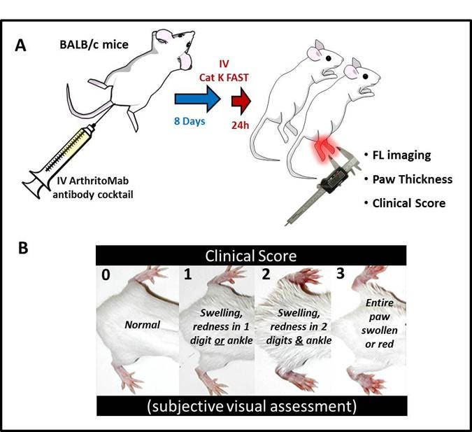
Monoclonal antibodies to collagen II can trigger an immune response in mice which provides a realistic model for rheumatoid arthritis. Eight days after treatment with the antibodies, the mouse exhibits pathology such as inflammation of the joint and synovium in addition to breakdown of the cartilage and bone. This model can be used in numerous mouse strains, including BALB/c which is typically resistant to other arthritis induction models (i.e. collagen-induced arthritis).
In Figure 2 (below), arthritis was induced in BALB/c mice and after 8 days the imaging probe IVISense CAT K 680 FAST was injected intravenously and the mouse was imaged 24 hours later. Fluorescent imaging results were compared to more traditional assessments of disease progression, such as clinical. As shown in Figure 3, 3D tomographic and 2D images were obtained from both arthritic and controlled mice and signal from fluorescence was compared to both paw thickness measurements and clinical score assessments.
Figure 1: Mouse antibody-induced arthritis. A) 5-week-old BALB/c mice are injected with 4 mg of ArthritoMab antibody cocktail followed 3 days later with LPS. After 8 days, IVISense CAT K 680 FAST is injected and the mouse is imaged 24 hour later, where fluorescent quantification is compared to paw thickness. B) Clinical score is typically assessed by visual assessment of paw swelling and redness.
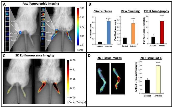
Figure 2: Assessment of arthritic mice. A) 3D tomographic images were obtained by FMT 2500 B) Paw thickness (vernier caliper), clinical observation score and quantification of IVISense CAT K FAST fluorescence from tomographic datasets C) 2D epifluorescence images acquired using FMT 2500 D) Ex vivo tissue analysis and 2D IVISense Cat K FAST epi-fluroescence imaging.
In Figure 3 below, the activatable probes IVISense Pan Cathepsin and IVISense MMP are used to assess protease activity in vivo, while the vascular imaging probe IVISense Vascular detects changes in vascular leak associated with arthritis
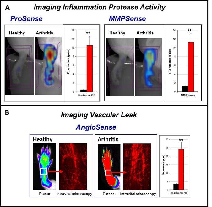
Figure 3: Activatable and Vascular probes used to image arthritis. A) The activatable probes IVISense Pan Cathepsin (formerly ProSense) and IVISense MMP (formerly MMPSense) are used to image protease activity associated with inflammation. B) The vascular probe IVISense Vascular (formerly AngioSense) is used to image vascular leak using both planar imaging and intravital microscopy measurements in both healthy and arthritic tissue.
In addition to in vivo and ex vivo measurement as shown above, probes can also be used to label cells and imaged using fluorescence microscopy. In Figure 4A below, human synovial fibroblasts were isolated from rheumatoid arthritis patients and labeled with IVISense CAT K 680 FAST. In Figure 4B rabbit synoviocytes (HIG-62 cell lines) were also labeled with the probe.
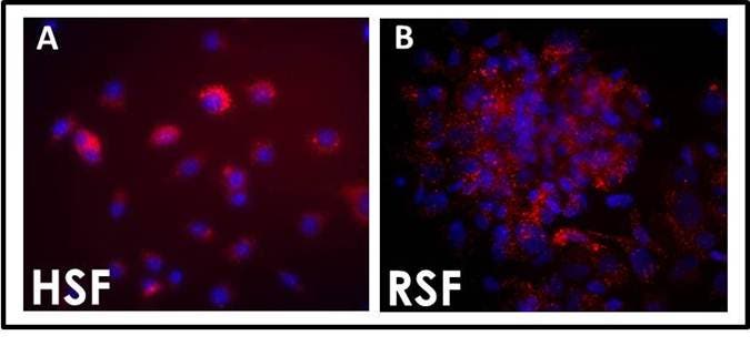
Figure 4: Uptake and activation of IVISense CAT K 680 FAST. Synovial fibroblasts isolated from human rheumatoid patient (A) or rabbit cell line (B). Cells were cultured with 1 mM IVISense CAT K FAST for 6 hours. Red: IVISense CAT K 680 FAST, Blue: DAPI nuclear stain; final magnification 40x.
Application notes and posters
- Poster: Imaging of Cathepsin K activity in rodent models of bone turnover and soft tissue calcification
- Poster: Development of a Fast Activating New Near Infrared-Labeled Probe for Detecting Cathepsin K Activity
The information provided above is solely for informational and research purposes only. Revvity assumes no liability or responsibility for any injuries, losses, or damages resulting from the use or misuse of the provided information, and Revvity assumes no liability for any outcomes resulting from the use or misuse of any recommendations. The information is provided on an "as is" basis without warranties of any kind. Users are responsible for determining the suitability of any recommendations for the user’s particular research. Any recommendations provided by Revvity should not be considered a substitute for a user’s own professional judgment. Users are solely responsible for complying with all relevant laws, regulations, and institutional animal care and use committee (IACUC) guidelines in their use of the information provided.