


Overview
Cathepsin (Cat) B is a lysosomal cysteine protease which is strongly overexpressed in cancers and premalignant lesions. Cathepsin B expression correlates with the invasiveness and metastatic capabilities of many tumors. In breast cancer, high expression levels of cathepsin B have been linked to highly aggressive tumors. Cathepsin B is also upregulated in a variety of inflammatory cells (including eosinophils, neutrophils, and macrophages). In the study of cardiovascular disease, expression levels of Cat B may provide an inflammation readout that indicates the potential vulnerability of atherosclerotic plaques.
IVISense™ Cat B 680 FAST and IVISense Cat B 750 FAST are fluorescent imaging probes developed for specific and rapid detection of tumors associated Cat B activity. They allow for the fast, non-invasive in vivo imaging of disease status and progression. They are optically silent upon injection and produce a fluorescent signal only after cleavage by Cathepsin B expressed in inflammatory cells and tumor cells. The probes consist of a pair of quenched fluorophores separated by a highly selective Cat B substrate. Upon cleavage by Cat B, the probe de-quenches and becomes highly fluorescent. In addition, they are conjugated to a pharmacokinetic modifier (PKM), which improves the pharmacokinetic profile. They are members of a family of activatable fluorescent imaging probes comprising this novel architecture, termed F.A.S.T. (Fluorescent Activatable Sensor Technology). This architecture offers higher target specific signal with reduced background and an improved pharmacokinetic profile with a broader range of early imaging time points.
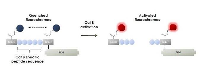
Figure 1: IVISense Cat B FAST fluorescent probes are composed of a Cat B-specific peptide flanked by two near-infrared (NIR) fluorochromes and a pharmacokinetic modifier (PKM) selected to provide optimal attributes for in vivo imaging. Upon Cat B cleavage of the substrate sequence the IVISense Cat B FAST fluorescent probes become highly fluorescent.
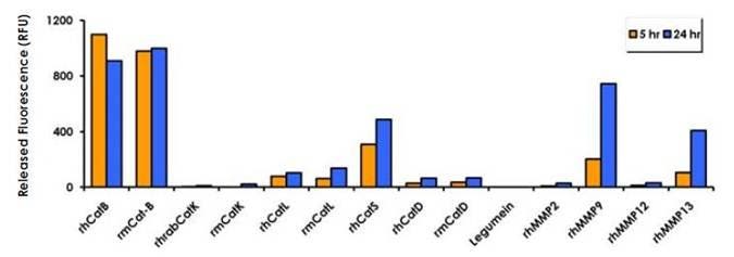
Figure 2: IVISense Cat B 750 FAST (0.5 mM) was cleaved in the presence of 0.05-0.1 mM activated human and mouse cathepsins B, K, L, S and D, legumain and MMPs 2, 9, 12, and 13 at an optimal pH and temperature. The fluorescence was read using a fluorescence microplate reader at 5 hours and 24 hours. IVISense Cat B 750 FAST is expected to be preferentially cleaved by Cat B in vivo, based on fast Cat B cleavage and short exposure profile in circulation (< 2 hours).
Products and catalog numbers
| Product | Catalog Number | Ex/Em wavelength (nm) | Molecular weight (g/mol) | Validated Experiments | Applications |
|---|---|---|---|---|---|
| IVISense Cat B 680 FAST | NEV11112 | 675/693 | 33,000 | In vivo/Ex vivo Flow cytometry In vitro microscopy |
Oncology Cardiovascular Inflammation Neurological diseases |
| IVISense Cat B 750 FAST | NEV11098 | 750/770 | 23,000 | In vivo/Ex vivo Flow cytometry In vitro microscopy |
Oncology Cardiovascular Inflammation Neurological diseases |
Using IVISense Cat B FAST fluorescent probe for in vivo/ex vivo studies
- For IVISense Cat B 680 FAST, we recommend intravenous injection and image 6-24 hours post injection. Some applications like imaging of atherosclerotic plaques may require a 4 nmol dose and may benefit from a later imaging time point.
- For IVISense Cat B 750 FAST, we recommend intravenous injection and imaging 6 hours post injection. Some applications like imaging of atherosclerotic plaques require an 8 nmol dose and may benefit from a later imaging time point.
- Instructions on setting up an in vivo mouse experiment with IVISense Cat B 680 FAST fluorescent probe
- Instructions on setting up an in vivo mouse experiment with IVISense Cat B 750 FAST fluorescent probe
| IVISense Cat B FAST | Route of Injection | Mouse Dose (25 g) | Rat Dose (250 g) | Blood t 1/2 | Tissue t 1/2 | Optimal imaging time | Optimal Re-injection Time (complete clearance) | Route of Metabolism/ background tissue | FMT and IVIS settings |
|---|---|---|---|---|---|---|---|---|---|
| IVISense Cat B 680 FAST | IV | 2 nmol; (4 nmol atherosclerosis) | 6-20 nmol | 5 min | 36 h | 6-24 h | 3 d | Salivary glands > liver, kidneys | FMT 680/700 IVIS 675/720 |
| IVISense Cat B 750 FAST | IV | 4 nmol; (8 nmol atherosclerosis) | 12 nmol | 5 min | 36 h | 6-24 h | 3 d | Salivary glands > liver, kidneys | FMT 750/770 IVIS 745/800 |
In Vivo/Ex Vivo Imaging
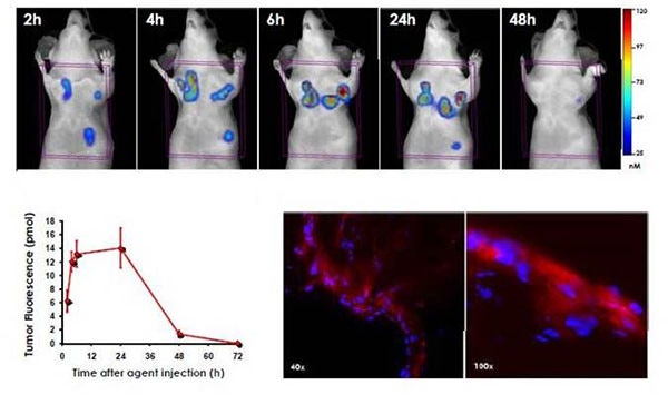
Figure 3: HT-29 tumor-bearing mice were injected intravenously with IVISense Cat B 750 FAST and imaged by fluorescence molecular tomography at 2, 4, 6, 24 and 48 hours. Panels show the tomographic images of a representative mouse at each time point. IVISense Cat B 750 FAST activation was quantified in pmols and signal was seen in as little as 2 hours. A plateau was reached between 6 and 24 hours which offers a large imaging window. Signal can easily be washed out by 48 hours. In the lower right panel, tumors were excised and snap-frozen after 6 hours and were visualized by fluorescence microscopy at 40x and 100x magnification. As shown here, IVISense Cat B 750 FAST is localized mostly at the tumor periphery but also inside the tumor.

Figure 4: HT-29 colorectal carcinoma cells were subcutaneously injected into the left and right mammary pad in 12 female athymic nu/nu mice. Eight days after implantation, when tumors had reached an average volume of 27 mm3, six animals received 170 mg/kg cyclophosphamide intraperitoneally and 40 mg cyclophosphamide/kg/day in their drinking water. Six remaining animals received no treatment. Tumor volume was monitored for ten days. Mice were then injected intravenously with IVISense Cat B 750 FAST in 100 μl of PBS and imaged 6 hours later using fluorescence molecular tomography imaging. A. This graph shows the tumor growth over a ten-day period with and without the cyclophosphamide treatment. B. The quantification of the tumor fluorescence (pmol) shows a significant increase in IVISense Cat B 750 FAST signal (72%) with cyclophosphamide treatment despite significantly smaller tumor volumes (46%) relative to control animals. C. Representative volume rendering projections of control and cyclophosphamide treated mice and anti-MAC3 immuno-fluorescent labeling (shown in green) in the ex vivo tumors, indicating increased TAM infiltration in cyclophosphamide treated animals at 40X magnification. DAPI staining is shown in blue.
Ex Vivo Imaging
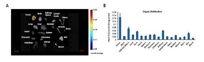
Figure 5: IVISense Cat B 750 FAST was intravenously injected into HT-29 tumor-bearing mice. Organs from these mice were excised at 6 hours after injection and imaged using fluorescence molecular tomography in reflectance mode. A) The 2D planar fluorescence image. B) Mean fluorescence intensity (FLU) of tumors and organs. IVISense Cat B 750 FAST activates primarily in tumors with less signal in other organs.
Frozen Tissue Protocol
We have validated IVISense Cat B 680 FAST for use with frozen tissue samples (IVISense Cat B 750 FAST has not been tested). Here is a brief protocol with a recommended concentration of probe to use:
- Freeze tumor/tissue (without probe) and section 5-10 µm by cryostat.
- Incubate with 1 µM IVISense Cat B 680 FAST at 37ºC for 10 min.
- Wash 2x with PBS.
- Mount with anti-fade reagent.
- Fluorescence microscopy filter: Cy5.5
Using IVISense Cat B FAST for in vitro studies
IVISense Cat B 680 FAST and IVISense Cat B 750 FAST can be used for detecting and monitoring Cat B activity in tumor and inflammatory cells lines.
Flow cytometry and in vitro microscopy
We have validated IVISense Cat B FAST for use with fluorescence microscopes and flow cytometers. Here is a brief protocol with a recommended concentration of probe to use:
- Culture cells in standard TC plate or chamber slide.
- Incubate cells with 1 µM IVISense Cat B 680 FAST or IVISense Cat B 750 FAST for 6-24 hours at 37°C.
- Wash 1x with PBS. For flow cytometry, detach and resuspend cells in PBS.
- Flow cytometry filter settings: 712/21 (IVISense Cat B 680 FAST), 780/60 (IVISense Cat B 750 FAST) Fluorescence micrsocopy filter: Cy 5.5 (IVISense Cat B 680 FAST), Cy5.5 or Cy7* (IVISense Cat B 750 FAST)
In Vitro Imaging
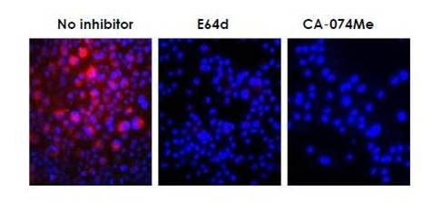
Figure 6: Raw 264.7 mouse macrophages were incubated with1 μM final concentration of IVISense Cat B 750 FAST. Cathepsin inhibitors (50 μM) were added 1 hour before addition of the probe. E64d cell-permeable cysteine/calpain inhibitor and CA-074Me specific cell-permeable cathepsin B inhibitor were used. Cells were then analyzed by fluorescence microscopy. IVISense Cat B 750 FAST is activated by RAW macrophages (shored), while the pan-cathepsin inhibitor E64d and the specific cathepsin B inhibitor CA-074Me significantly reduce the activation.
FAQs
Q. Can I use IVISense Cat B FAST probes in humans?
A. No, IVISense Cat B FAST probes are intended for animal research use only, are not intended for use in diagnostic procedures and are not intended for use in humans.
Q. Is it possible to obtain a functionalized version of IVISense Cat B 680 FAST for covalent attachment to a lipid (e.g., a carboxylic acid)?
A. We don’t currently have any IVISense Cat B probes that can attach to a lipid
For research use only. Not for use in diagnostic procedures. The information provided above is solely for informational and research purposes only. The information does not constitute medical advice and must not be used or interpreted as such. Consult a qualified veterinarian or researcher for specific guidance or use information. Revvity assumes no liability or responsibility for any injuries, losses, or damages resulting from the use or misuse of the provided information, and Revvity assumes no liability for any outcomes resulting from the use or misuse of any recommendations. The information is provided on an "as is" basis without warranties of any kind. Users are responsible for determining the suitability of any recommendations for the user’s particular research. Any recommendations provided by Revvity should not be considered a substitute for a user’s own professional judgment. Users are solely responsible for complying with all relevant laws, regulations, and institutional animal care and use committee (IACUC) guidelines in their use of the information provided.