


Overview
Cathepsin K (Cat K) is a lysosomal cysteine protease expressed in osteoclasts, chondrocytes, and synovial fibroblasts and is involved in bone resorption and collagen degradation. Women with osteoporosis, breast cancer and bone metastases have high levels of Cat K and therefore, Cat K expression is a good marker for these diseases. In addition, researchers have been looking for inhibitors of Cat K for potential therapeutics. Cat K is also expressed by a broad range of other cell types including ovarian cells, colonic tissue, alveolar and bronchial epithelial cells, synovial fibroblasts, and macrophages.
IVISense™ Cat K 680 FAST fluorescent probe was developed as an imaging tool for monitoring and quantifying Cat K activity. IVISense Cat K 680 FAST is an activatable probe that is optically silent upon injection and only produces fluorescent signal after cleavage by Cathepsin K. It contains a human Cat K-cleavable sequence with a pair of self-quenching NIRF fluorochomes. In addition, a pharmacokinetic modifier was added to confer increased blood half-life. Disease status and progression can easily be monitored in vivo with this non-invasive probe. IVISense Cat K 680 FAST offers higher target specific signal with reduced background while also reducing the optimal imaging time after injection.
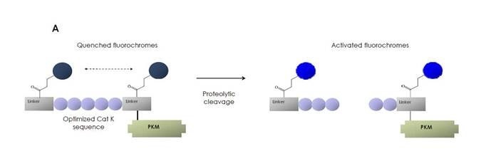
Figure 1: IVISense Cat K 680 FAST is comprised of a Cat K-specific peptide sequence flanked by two near-infrared (NIR) fluorochromes. It includes a pharmacokinetic modifier (PKM) selected to confer increased blood half-life. The probe is quenched (no fluorescence) until cleavage by Cat K when it becomes highly fluorescent.
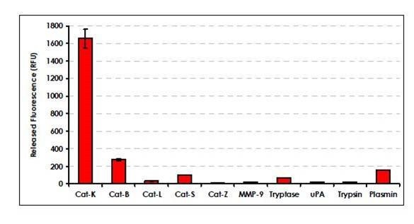
Figure 2: IVISense Cat K 680 FAST is selectively cleaved by Cat K over a variety of other enzymes. IVISense Cat K 680 FAST (0.5 µM) was incubated with a variety of enzymes and the fluorescence released was measured after 5 hours.
Products and catalog numbers
| Product | Catalog Number | Ex/Em wavelength (nm) | Molecular weight (g/mol) | Validated Experiments | Applications |
|---|---|---|---|---|---|
| IVISense Cat K 680 FAST | NEV11000 | 674/692 | 8500 | In vivo/Ex vivo | Oncology |
| Flow cytometry | Atherosclerosis | ||||
| In vitro microscopy | Arthritis | ||||
| Osteoporosis | |||||
| Lung remodeling | |||||
| Obesity |
Using IVISense Cat K 680 FAST probe for in vivo/ex vivo studies
The recommended procedure for in vivo imaging with IVISense Cat K 680 FAST fluorescent probe is administration via intravenous injection and imaging 6 hours post injection. Optimal imaging time is 6-24 hours after injection for IVISense Cat K 680 FAST depending on the model.
- Instructions on setting up an in vivo mouse experiment using IVISense Cat K 680 FAST fluorescent probe
| Route of Injection | Mouse Dose (25 g) | Rat Dose (250 g) | Blood t 1/2 | Tissue t 1/2 | Optimal imaging time | Optimal Re-injection Time (complete clearance) | Route of Metabolism/ background tissue | FMT and IVIS settings |
|---|---|---|---|---|---|---|---|---|
| IV | 2 nmol | 6-20 nmol | 30 min | 36 h | 6-24 h | 3 d | Kidney > liver | FMT 680/700 |
| IVIS 675/720 |
In Vivo/Ex vivo Imaging
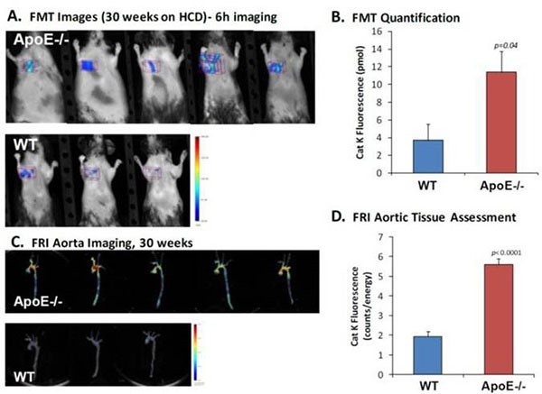
Figure 3: ApoE-/- mice were placed on a high-cholesterol diet for 30 weeks (n =5). These mice and the control mice (n = 3) were injected with 8 nmols per mouse IVISense Cat K 680 FAST and imaged by FMT 2500. A) The representative FMT 2500 images from control and apoE-/- mice. Images were taken 6 hours following injection and tissues were collected from perfused animals immediately. B) The quantification of fluorescence molecular tomography aortic arch region fluorescence from non-invasive imaging datasets (used optimal threshold to minimize background fluorescence). C) The corresponding ex vivo FRI images of aortic tissue (from the same mice). D) The corresponding ex vivo FRI analysis of aortic arch tissue, focusing the quantification on the aortic arch region only.
Using IVISense Cat K 680 FAST probe for in vitro studies
IVISense Cat K 680 FAST can be used for detecting and monitoring Cat K activity in synovial fibroblasts from rheumatoid arthritis (RA) and bone marrow-derived macrophages.
Flow cytometry and in vitro microscopy
We have validated IVISense Cat K 680 FAST for use with fluorescence microscopes and flow cytometers. Here is a brief protocol with a recommended concentration of probe to use:
- Culture cells in standard TC plate or chamber slide.
- Incubate cells with 0.5 µM IVISense Cat K 680 FAST for 6 hours at 37 °C.
- Wash 1x with PBS. For flow cytometry, detach and resuspend cells in PBS.
- Flow cytometry filter settings: 712/21 Fluorescence micrsocopy filter: Cy5.5
In Vitro Imaging
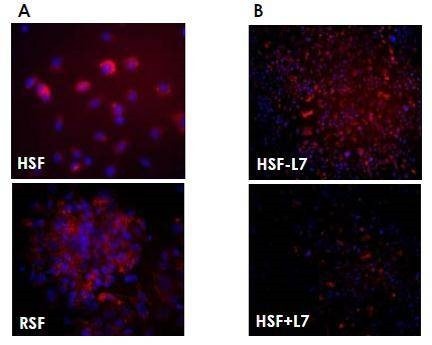
Figure 4: A) Cells (human synovial fibroblasts (HSF) from a patient with RA (upper) and rabbit synoviocytes (HIG-62) (lower)) were cultured in the presence of 1 µM IVISense Cat K 680 FAST for 6 hours. IVISense Cat K 680 FAST activation is shown in red and DAPI nuclear stain is shown in blue and the final magnification is 40x. B) As a control, HSF cells were pre-incubated in the absence (top) or presence (lower panel) of a specific Cathepsin K inhibitor (L7, 200 nM) for 1 hour before addition of the IVISense Cat K 680 FAST. The cells were cultured for an additional 6 hours and as shown in these panels, IVISense Cat K 680 FAST activation is inhibited. The final magnification for these panels is 20x.
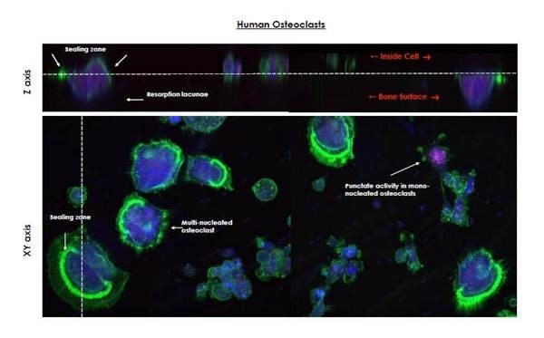
Figure 5: Human osteoclasts were cultured on bovine cortical bone for seven days. IVISense Cat K 680 FAST (1 µM) was added for 24 hours and then the cells were visualized by confocal microscopy. Using activated IVISense Cat K 680 FAST (blue), Cathepsin K activity was easily detected in the resorption lacunae and intracellularly in both mononuclear multinucleated cells. Acidic vesicles in lysozomes were stained using Lysotracker (shown in red) and osteoclast actin rings were visualized using FITC-phalloidin (shown in green).
Application notes and posters
- Poster: Imaging of Cathepsin K activity in rodent models of bone turnover and soft tissue calcification
- Poster: Development of a Fast Activating New Near Infrared-Labeled Probe for Detecting Cathepsin K Activity
FAQs
Q. Can I use IVISense Cat K 680 FAST in humans?
A. No, IVISense Cat K 680 FAST is intended for animal research use only, is not intended for use in diagnostic procedures and is not intended for use in humans.
Q. What is the difference between IVISense Cat K 680 FAST and the OsteoSense probes?
A. IVISense Cat K is useful in pathological models, such as Osteoporosis, where bone resorption and soft tissue calcification occurs. IVISense Osteo probe, on the other hand, specifically targets areas of bone growth and loss. It does this by binding to hydroxyapatite, a major mineral product of osteoblasts and, thus, marker of osteoblast activity.
For research use only. Not for use in diagnostic procedures.