


Traditional mouse models of cancer rely primarily on ex vivo measurements of disease morphology and histologic analysis for the assessment of tumors. These measurements of disease may be distant from the actual biological targets of interest and can be time consuming, expensive, and impractical. By using NIR (Near-Infrared) imaging probes in combination with optical in vivo systems, the biological processes that change with disease progression and therapeutic response can be visualized non-invasively over time.
IVISense™ Pan Cathepsin fluorescent probes (formerly ProSense) were developed to specifically look at the expression and activity of key disease associated proteases. The probes are optically silent until in the presence of these proteases and can be used to easily monitor the activity of these proteases in real time. Revvity offers three different IVISense Pan Cathepsin activatable fluorescent probes. IVISense Pan Cathepsin 680 is activated by Cathepsin B, L, S and Plasmin. IVISense Pan Cathepsin 750 FAST is activated by Cathepsin B, L, S, K, V and D. IVISense Pan Cathepsin 750 is activated by Cathepsin B, L, S and Plasmin.
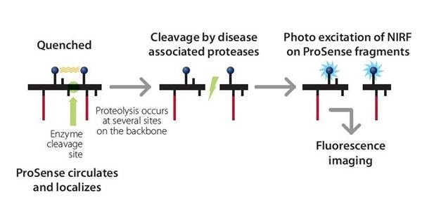
Figure 1: Mechanism of IVISense Pan Cathepsin (formerly ProSense) activation. The IVISense Pan Cathepsin probes are comprised of a pair of quenched fluorophore separated by a cleavable linker. Upon cleavage of the linker by the specific disease-related proteases, the probe becomes highly fluorescent. Therefore, the fluorescent signal is directly proportional to the expression and activity levels of the proteases.
Products and catalog numbers
| Product | Catalog Number | Ex/Em wavelength (nm) | Molecular weight (g/mol) | Validated Experiments | Applications |
|---|---|---|---|---|---|
| IVISense Pan Cathepsin 680 | NEV10003 | 680/700 | 400,000 | In vivo/Ex vivo | Oncology |
| Flow cytometry | Arthritis | ||||
| In vitro microscopy | |||||
| IVISense Pan Cathepsin 750 FAST | NEV11171 | 750/780 | 400,000 | In vivo/Ex vivo | Oncology |
| Flow cytometry | Arthritis | ||||
| In vitro microscopy | |||||
| IVISense Pan Cathepsin 750 | NEV10001EX | 750/770 | 450,000 | In vivo/Ex vivo | Oncology |
| Flow cytometry | Inflammation | ||||
| In vitro microscopy |
Using IVISense Pan Cathepsin fluorescent probes for in vivo studies
The recommended procedure for in vivo imaging with IVISense Pan Cathepsin fluorescent probes is administration via tail vein injection and imaging 24 hours post injection.
- Imaging in Arthritis: IVISense Pan Cathepsin fluorescent probes can be used as a marker for disease progression and therapeutic response in animal models of arthritis.
- Imaging in Oncology: IVISense Pan Cathepsin fluorescent probes can be used as a marker for disease progression in animal tumor models.
-
Pulminary Disease: IVISense Pan Cathepsin fluorescent probes to assess cathepsin activity in activated neutrophils.
Instructions on setting up an in vivo mouse imaging experiment with:
- IVISense Pan Cathepsin 680 fluorescent probe
- IVISense Pan Cathepsin 750 FAST fluorescent
- probe IVISense Pan Cathepsin 750 fluorescent probe
| Product | Route of Injection | Mouse Dose (25 g) | Rat Dose (250 g) | Blood t 1/2 | Tissue t 1/2 | Optimal imaging time | Optimal Re-injection Time (complete clearance) | Route of Metabolism/ background tissue | FMT and IVIS settings |
|---|---|---|---|---|---|---|---|---|---|
| IVISense Pan Cathepsin 680 | IV | 2 nmol | 6-20 nmol | 12 h | 72 h | 24 h (24-48) | 6-7 d | Liver | FMT 680/700 |
| IVIS 675/720 | |||||||||
| IVISense Pan Cathepsin 750 FAST | IV | 2 nmol | 6-20 nmol | 24 h | 72 h | 24 h | 6-7 d | Low liver, intestine | FMT 750/770 |
| IVIS 745/800 | |||||||||
| IVISense Pan Cathepsin 750 | IV | 4 nmol | 12-40 nmol | 30 min | 36 h | 6-24 h | 3 d | Low liver, bladder | FMT 750/770 |
| IVIS 745/800 |
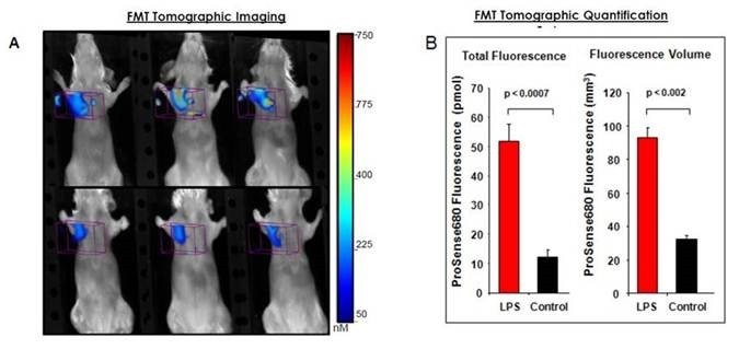
Figure 2: A) Precise fluorescence localization of activated IVISense Pan Cathepsin 680 is shown by FMT imaging. The non-invasive tomographic imaging is able to detect disease-related protease activity and accurate quantification of the internal distribution of this fluorescence in the lung. B) Fluorescence molecular tomography measured a 5-fold increase in IVISense Pan Cathepsin 680 fluorescence in the lungs of chronic obstructive pulmonary disease (COPD, asthma) mice (> 55 pmol/lung) as compared to those of control mice (< 10-15 pmol/lung). Tomography software precisely quantified the volume of the fluorescence within the lung regions of the mice to provide a measure of affected lung volume, showing approximately a 3-fold increase in COPD animals as compared to controls (~100 mm3 in asthma vs 30 mm3 in controls). Both the total fluorescence quantification (pmol) and the volume measurement (mm3) show high statistical differences in comparing asthma and control mice (p<0.0007 and p<0.002, respectively).
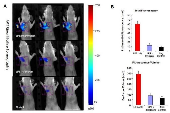
Figure 3: Rolipram (a PDE4 inhibitor) has been proven as an anti-inflammatory drug. To evaluate whether the effect of a PDE4 inhibitor on COPD treatment could be detected and measured non-invasively using fluorescence molecular tomography, mice were treated with Rolipram (2 mg/kg given intraperitoneally 24, 12, and 0.5 hours prior to intranasal lipopolysaccharide treatment (LPS), which induces inflammation). Control mice received injections of PBS. All mice were injected with IVISense Pan Cathepsin 680 4hours after LPS treatment. A) Molecular tomography showed high fluorescence signal in untreated asthmatic mice compared to Rolipram-treated mice. B) The quantification of the therapeutic response in treated LPS-induced lung inflammation showed 92% inhibition of total IVISense Pan Cathepsin 680 signal and 86% inhibition of fluorescence volume.
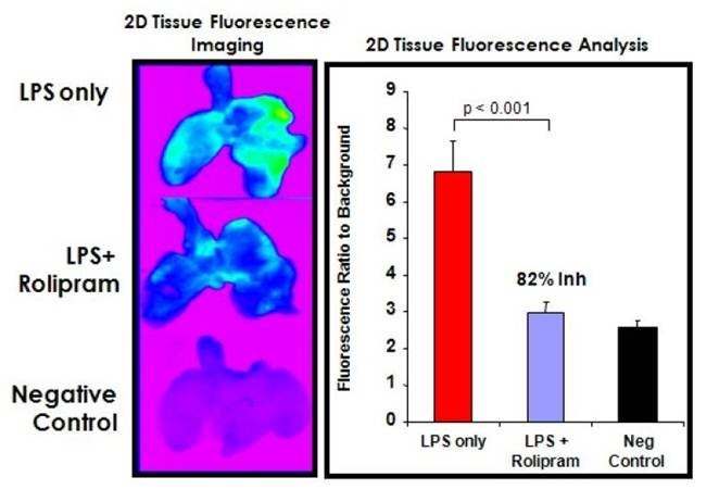
Figure 4: Tissue 2D imaging of IVISense Pan Cathepsin 680 fluorescence was measured ex vivo to verify the dramatic effects of Rolipram treatment on LPS lung inflammation and on non-invasive fluorescence tomography quantification. Image analysis to generate tissue fluorescence ratios to background yielded a highly significant (p < 0.001) inhibition of IVISense Pan Cathepsin 680 tissue fluorescence by Rolipram treatment.
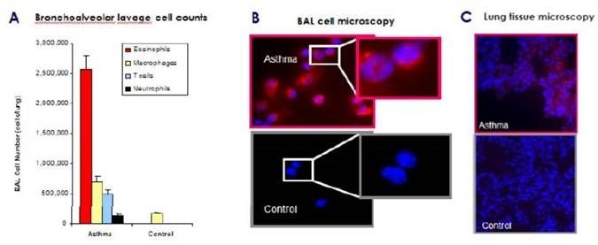
Figure 5: BAL cell differential analysis indicates the expected recruitment eosinophils (> 60% of the total cells) infiltrating the airways of asthmatic mice and no detectable eosinophils in control mice. A) Fluorescent microscopy of the BAL cells isolated from asthmatic and control mice that received intravenous IVISense Pan Cathepsin 680 injection. This data shows IVISense Pan Cathepsin 680 fluorescence is predominantly activated by eosinophils cells in the airways. B) BAL mononuclear cells in the same mice showed little or no fluorescence, whereas BAL cells from control mice (predominantly macrophages) showed minimal fluorescence. C) Tissue sections from the lungs of asthmatic mice demonstrated multiple areas of fluorescence localized to the lung parenchyma. Minimal fluorescence is detectable in the control lungs.
Using IVISense Pan Cathepsin fluorescent probes for in vitro studies
IVISense Pan Cathepsin fluorescent probes have been validated for use with tumor and inflammatory cells.
Flow cytometry and in vitro microscopy
We have validated IVISense Pan Cathepsin fluorescent probes for use with fluorescence microscopes and flow cytometers. Here is a brief protocol with a recommended concentration of probe to use:
- Culture cells in standard TC plate or chamber slide.
- Incubate cells with 1 μM IVISense Pan Cathepsin 680, 1-2 μM IVISense Pan Cathepsin 750, or 1 μM IVISense Pan Cathepsin 750 FAST for 6-24 hours at 37 °C.
- Wash 1x with PBS. For flow cytometry, detach and resuspend cells in PBS.
- Flow cytometry filter settings: 712/21 (IVISense Pan Cathepsin 680), 780/60 (IVISense Pan Cathepsin 750 or IVISense Pan Cathepsin 750 FAST). Fluorescence micrsocopy filter: Cy5.5 (IVISense Pan Cathepsin 680), Cy5.5 or Cy7* (IVISense Pan Cathepsin 750 or IVISense Pan Cathepsin 750 FAST).
In Vitro Imaging
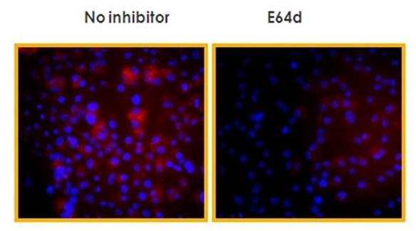
Figure 6: Raw 264.7 mouse macrophages were incubated with IVISense Pan Cathepsin 750 FAST (1 μM final concentration). The cell-permeable cysteine/calpain inhibitor was added 1 hour before addition of the probe. Cells were analyzed by fluorescence microscopy. IVISense Pan Cathepsin 750 FAST is activated by RAW macrophages. The pan-cathepsin inhibitor E64d significantly reduces the activation.
Application notes and posters
- Application Note: Quantitative Pre-clinical Fluorescence Imaging of Cancer Metastasis to the Lung and Response to Therapy
- Application Note: Noninvasive, In Vivo Quantitation of Asthma Severity using Fluorescence Molecular Tomography
- Application Note: Non-Invasive Quantitative In Vivo Imaging of Atherosclerosis Disease Progression and Treatment Response in ApoE Deficient Mice using Fluorescence Molecular Tomography and NIR Fluorescent Pre-clinical Imaging Agents
FAQs
Q. Can IVISense Pan Cathepsin fluorescent probes be used in humans?
A. No, IVISense Pan Cathepsin fluorescent probes are intended for animal research only, are not intended for use in diagnostic procedures and not intended for use in humans.
Q. What is the difference between IVISense Pan Cathepsin 750 and IVISense Pan Cathepsin 750 FAST?
A. IVISense Pan Cathepsin 750 FAST is one of our F.A.S.T. (Fluorescent Activatable Sensor Technology) probes that confers an improved pharmacokinetic profile with earlier imaging time points. It is activated by Cathepsin B, L, S, K, V and D and is not cleaved by trypsin or plasmin. IVISense Pan Cathepsin 750 is activated by Cathepsin B, L, S and Plasmin. IVISense Pan Cathepsin 750 has a much faster clearance time (see above table).
For research use only. Not for use in diagnostic procedures.
The information provided above is solely for informational and research purposes only. The information does not constitute medical advice and must not be used or interpreted as such. Consult a qualified veterinarian or researcher for specific guidance on any injection techniques or other animal procedures. Revvity assumes no liability or responsibility for any injuries, losses, or damages resulting from the use or misuse of the provided information. The information is provided on an "as is" basis without warranties of any kind. Users are solely responsible for complying with all relevant laws, regulations, and institutional animal care and use committee (IACUC) guidelines in their use of the information provided.