


Overview
The vascular system is made up of the vessels that carry blood and lymph through the body. It supplies all organs and tissues of the body with oxygen and nutrients, removal of waste products, fluid balance, and other functions. Changes in the vascular system may occur in a variety of different cancers and inflammatory states. In vivo imaging enables studying these changes.
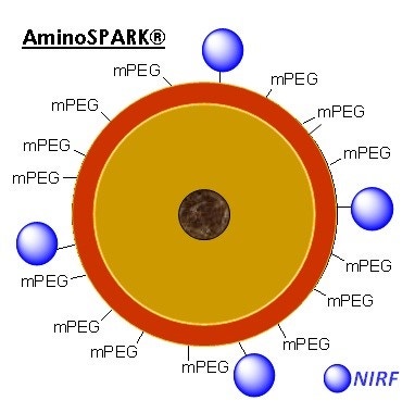
IVISense™ Vascular NP fluorescent nanoparticles (formerly AngioSPARK) are a family of highly-fluorescent near infrared nanoparticles specifically designed for in vivo imaging. They contain an iron oxide core that is coated to specifically produce a functionalized biocompatible product. IVISense Vascular NP 680 fluorescent nanoparticles comprises pegylated fluorescent nanoparticles that remain localized in the vasculature for extended periods of time and enable imaging of blood vessels and angiogenesis.
Products
| Product | Catalog Number | Ex/Em wavelength (nm) | Nanoparticle size | Validated Experiments | Applications | Storage and Stability |
|---|---|---|---|---|---|---|
| IVISense Vascular NP 680 | NEV10149 | 673 ± 5 nm | 20 – 50 nm | Intravital microscopy imaging administration via tail vein injection and imaging | Vascularity in cancer and inflammation | Store at 2-8 °C protected from light. Stable up to 12 months. |
| 690 ± 5 nm | ||||||
| IVISense Vascular NP 750 | NEV10150 | 750 ± 5 nm | 20 – 50 nm | Intravital microscopy imaging administration via tail vein injection and imaging | Vascularity in cancer and inflammation | Store at 2-8 °C protected from light. Stable up to 12 months. |
| 775 ± 5 nm |
Using IVISense Vascular NP probe for in vivo studies
The recommended procedure for in vivo imaging with IVISense Vascular NP is administration via tail vein injection and imaging 0-4 hours post injection.
Imaging in Oncology: IVISense Vascular NP can be used to study angiogenesis, as a marker for blood vessel density, in animal tumor models.
Imaging in Arthritis: IVISense Vascular NP can be used to characterize vascular changes and therapeutic responses associated with animal models of arthritis.
| Product | Route of Injection | Mouse Dose (25 g) | Rat Dose (250 g) | Blood t 1/2 | Tissue t 1/2 | Optimal imaging time | Optimal Re-injection Time (complete clearance) | Route of Metabolism/ background tissue | FMT and IVIS settings |
|---|---|---|---|---|---|---|---|---|---|
| IVISense Vascular NP 680 | IV | 100 uL | 300 - 1000 uL | 20 h | >100 h | 24 h | Pre-image subtraction | Long term tissue accumulation | - FMT 680/700 |
| - IVIS 675/720 | |||||||||
| IVISense Vascular NP 750 | IV | 100 uL | 300 - 1000 uL | 20 h | >100 h | 24 h | Pre-image subtraction | Long term tissue accumulation | - FMT 750/770 |
| - IVIS 745/800 |
In Vivo Imaging
Determining optimal tumor imaging timepoints for IVISense Vascular NP.
To determine the optimal imaging point in time relative to probe injection, we imaged 4T1 tumor-bearing mice at different times post-IVISense Vascular NP 680 injection (Figure 2). Whole body images at 24h post-injection (Fig 2A) reveal signal throughout the body (including bladder, intestines, liver, and heart) as well as in the tumor. Careful FMT assessment of the tumor regions showed a mostly vascular signal from 5 minutes to 3 h. The maximal tumor signal over background was seen at 24 h, with signal distributed throughout the tumors at 24-48 h. At later time points (72-96 h) signal appears to clear from the center of the tumors and localizes predominantly in the tumor margins. Both FMT and planar imaging can detect differences as compared to control sites. FMT would offer the additional advantage of the detection of deep tissue tumors undetectable at the surface by 2D planar imaging.
Figure 2. Imaging of tumors with IVISense Vascular NP. Fluorescence molecular tomograrphy and planar images show the patterns of fluorescence that occur in animals bearing tumor masses on the upper mammary fat pads. A) Whole body signal using fluorescence tomography. B) Tomographic imaging showing only tumor region fluorescence over time. Inset panels represent surface fluorescence detected by planar imaging.
For research use only. Not for use in diagnostic procedures. The information provided above is solely for informational and research purposes only. The information does not constitute medical advice and must not be used or interpreted as such. Consult a qualified veterinarian or researcher for specific guidance or use information. Revvity assumes no liability or responsibility for any injuries, losses, or damages resulting from the use or misuse of the provided information, and Revvity assumes no liability for any outcomes resulting from the use or misuse of any recommendations. The information is provided on an "as is" basis without warranties of any kind. Users are responsible for determining the suitability of any recommendations for the user’s particular research. Any recommendations provided by Revvity should not be considered a substitute for a user’s own professional judgment. Users are solely responsible for complying with all relevant laws, regulations, and institutional animal care and use committee (IACUC) guidelines in their use of the information provided.