Menu
US
US

Targeted Protein Degradation

Protein homeostasis relies on the ubiquitin-proteasome system (UPS) and the autophagy-lysosomal system (ALS) to degrade proteins and clear misfolded polypeptides and organelles.

Targeted protein degradation harnesses these pathways to overcome drug resistance and selectively target “undruggable” proteins, offering therapeutic possibilities beyond traditional drugs. PROTAC®s and molecular glues are leading modalities, enabling precise protein degradation to address complex diseases.

Molecular glues enhance interactions between target proteins and E3 ubiquitin ligases, driving degradation and providing solutions for challenging targets.
 

image-1-protac-mechanism-800-px

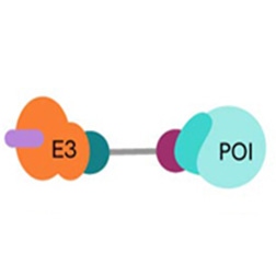
PROTACs induce targeted protein degradation by serving as a bridge between a Protein of Interest (POI) and an E3 ligase. The resulting ternary complex catalyzes polyubiquitination of the target protein, tagging it for degradation by the proteasome.

Technologies to advance your research

AlphaLISA™ SureFire® Ultra™ and HTRF™ Phospho/Total Protein Detection: No-wash assay kits offer a powerful alternative to traditional methods like western blotting, streamlining workflows and boosting throughput. These kits enable precise, quantitative measurement of phospho and total protein levels as well as protein-protein interactions, all without the need for extensive washing steps. Our cell-based phospho and total kits are designed to measure endogenous, untagged protein expression levels across a wide range of biological samples, providing valuable insights into native protein dynamics in real-time. Additionally, they are highly effective in assessing the degradation of target proteins induced by degraders, empowering researchers to monitor the effects of degraders with enhanced accuracy and efficiency.

AlphaLISA and HTRF E3 Ligase Binding Kits: The study of E3 ligases in drug discovery, particularly in PROTAC (PROteolysis TArgeting Chimeras) development, are crucial due to the diverse role of E3 ligases in tagging target proteins for degradation. These ligases play a central part in maintaining protein homeostasis and represent attractive therapeutic targets. In this context, AlphaLISA and HTRF E3 ligase binding kits offer robust, sensitive methods to analyze and quantify E3 ligase interactions with small molecules or PROTACs, streamlining the drug discovery process.
 

Explore more on degraders, mechanisms, and our assay offerings below.

Targeted Protein Degradation
Imagesection4-252x252

Is the target protein effectively degraded?

Poly-ubiquitination of the POI is followed by its recruitment by the proteasome and its proteolytic degradation. When the degradation rate of a target protein is faster than the rate at which it is being expressed, its net intracellular level decreases. If the target protein is also a pharmacological target, the effects of this decrease can be observed through downstream pathway effects. A reliable assessment of degradation is therefore an indirect measure of a PROTAC molecule’s binding efficacy and ultimately, its usefulness as a degrader.

Poly-ubiquitination of the POI is followed by its recruitment by the proteasome and its proteolytic degradation. When the degradation rate of a target protein is faster than the rate at which it is being expressed, its net intracellular level decreases. If the target protein is also a pharmacological target, the effects of this decrease can be observed through downstream pathway effects. A reliable assessment of degradation is therefore an indirect measure of a PROTAC molecule’s binding efficacy and ultimately, its usefulness as a degrader.

We offer a wide range of detection solutions for assessing protein degradation which ensure selectivity, sensitivity, and reproducibility and are ideal for replacing Western Blotting. Our AlphaLISA SureFire® Ultra™ total protein kits and HTRF total protein kits are alternatives to Western Blots for monitoring protein expression levels with precision. Our vast phospho protein kit portfolio offers a way to characterize the mechanism of action, to measure the functionality or effect of a degrader downstream.

image-11-alphalisa-htrf-protein-degradation-assays


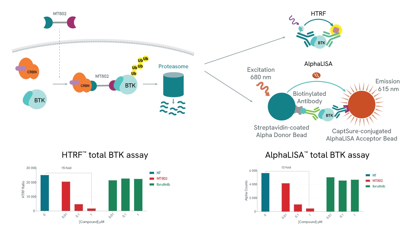
Protein degradation can be easily assessed using our total protein assays. In both examples (HTRF and AlphaLISA), use of a BTK-specific PROTAC, MT-802 leads to a drastic reduction of the detected levels of BTK. As expected, BTK levels are not affected by increasing concentrations of Ibrutinib, a BTK inhibitor.
 

image-htrf-egfr


PROTAC targeting EGFR assessed with total and phospho kits.
 

image-brd4-atplite


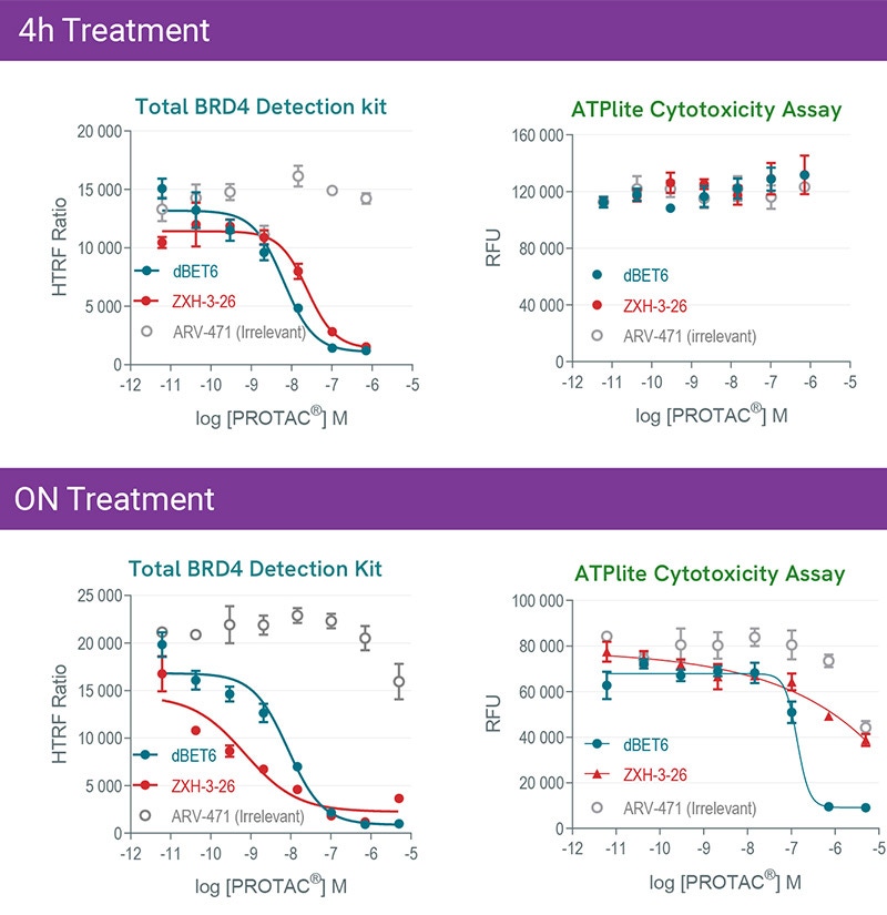
Kinetics of dBET6 and ZXH-3-26 PROTAC®s mediated degradation and cytotoxicity effects. HeLa cells were treated 4h or overnight with increasing concentrations of PROTAC®s. Experiments were carried out following instruction of each kit to monitor BRD4 expression level with HTRF total BRD4 kit, and Cytotoxicity with ATPlite kit.
 

image-cMyc


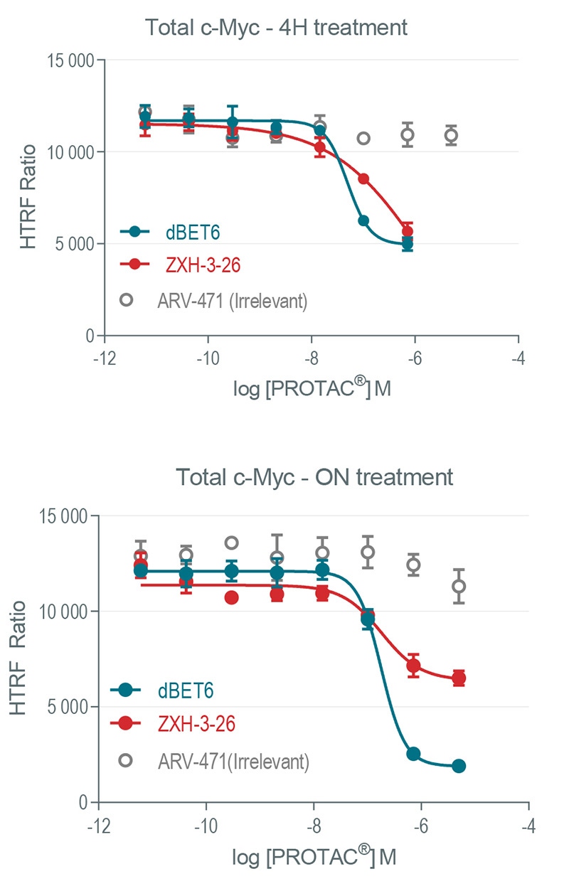
Modulation of c-Myc expression as a downstream BRD4 readout after PROTAC® treatment. HeLa cells were treated with increasing concentrations of dBET6 (pan BET family), ZXH-3-26 (selective BRD4 PROTAC®) as well as ARV-471 irrelevant PROTAC®) for 4h or overnight.
 

Imagesection1-252x252

Is the degrader binding to the protein of interest?

Affinity for the POI is not the only consideration when designing a PROTAC molecule. As a therapeutic agent, the first three barriers it must overcome in an in vivo environment are chemical stability, solubility and membrane permeability.

Affinity for the POI is not the only consideration when designing a PROTAC molecule. As a therapeutic agent, the first three barriers it must overcome in an in vivo environment are chemical stability, solubility and membrane permeability.

Once in the cellular environment, the PROTAC first forms a binary complex with either the POI or the specific E3 ligase. Successful engagement of both binding partners signals the formation of the ternary complex.

Efficient and fast compound optimization is of paramount importance in the drug discovery process. Revvity offers many tools that can be used in PROTAC characterization studies.

Kinase-binding assays

If your POI is a kinase, Revvity’s biochemical offering includes three kinase activity assay kits that allow the measuring of the binding affinity of your PROTAC binder and are compatible with 80% of the human kinome. The assays are based on the competitive dissociation of a known kinase-binding inhibitor by the compound under study.
 

image-3-kinase-binding-assays-800px


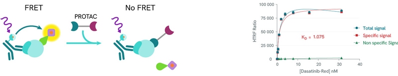
HTRF® kinase binding kits from Revvity are sandwich immunoassays based on Fluorescence Resonance Energy Transfer (FRET) between a FRET donor (a conjugated anti-6HIS, anti-GST or anti-biotin antibody) and a fluorescent acceptor tracer. Competitive displacement of the tracer by the PROTAC reduces the fluorescent signal allowing the calculation of a dissociation constant.

Imagesection2-252x25

Is the degrader binding to the E3 Ligase?

With only 2 E1 enzymes and 30-50 E2 enzymes encoded in the human genome, E3 ligases exhibit remarkable diversity, with over 600 genes. This variety is key in PROTAC (PROteolysis TArgeting Chimeras) design, as different ligases can be recruited to target specific proteins for degradation. A common E3 ligase in PROTAC design is Cereblon (CRBN), which forms an E3 ligase complex with DDB1 (DNA Damage Binding protein 1), Cullin-4A (CUL4A), and ROC1 (Regulator of Cullins 1).

With only 2 E1 enzymes and 30-50 E2 enzymes encoded in the human genome, E3 ligases exhibit remarkable diversity, with over 600 genes. This variety is key in PROTAC (PROteolysis TArgeting Chimeras) design, as different ligases can be recruited to target specific proteins for degradation. A common E3 ligase in PROTAC design is Cereblon (CRBN), which forms an E3 ligase complex with DDB1 (DNA Damage Binding protein 1), Cullin-4A (CUL4A), and ROC1 (Regulator of Cullins 1).

image-5-ubiquitin-e3-ligase-role-800p


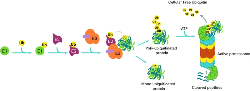
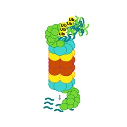
The process of ubiquitination can be sequentially repeated resulting in poly-ubiquitinated proteins which are then directed to the proteasome for degradation.

Assessing PROTAC binding to E3 ligases

With only 2 E1 enzymes and 30-50 E2 enzymes encoded in the human genome, E3 ligases exhibit remarkable diversity, with over 600 genes. This variety is key in PROTAC (PROteolysis TArgeting Chimeras) design, as different ligases can be recruited to target specific proteins for degradation. A common E3 ligase in PROTAC design is Cereblon (CRBN), which forms an E3 ligase complex with DDB1 (DNA Damage Binding protein 1), Cullin-4A (CUL4A), and ROC1 (Regulator of Cullins 1).

Revvity supports PROTAC development with a suite of AlphaLISA and HTRF E3 ligase binding kits, which facilitate precise binding studies. Sample data generated using the AlphaLISA Cereblon Binding Kit illustrate the competitive binding of various molecules to Cereblon. For example, the PROTAC dBRD9 and the IMiDs (Immunomodulatory imide drugs), Pomalidomide and Lenalidomide, all competitively bind to Cereblon, displacing a biotinylated Cereblon ligand and generating dose-response inhibition curves that allow IC50 determination. However, VH032, a ligand that recruits the VHL (Von Hippel-Lindau) E3 ligase rather than Cereblon, does not displace the Cereblon ligand.

Similarly, HTRF assays demonstrate that the PROTAC MT-802 and Pomalidomide compete with a fluorescently labeled Thalidomide ligand for Cereblon binding, whereas VH032 (specific to VHL) does not. MT-802, a PROTAC targeting BTK (Bruton’s Tyrosine Kinase) for degradation, and Pomalidomide produce dose-dependent inhibition, enabling IC50 calculations that align with established literature values. These kits offer valuable insights into PROTAC-E3 ligase interactions and support precise PROTAC design for targeted protein degradation.

image-6-alphalisa-htrf-cereblon-assays


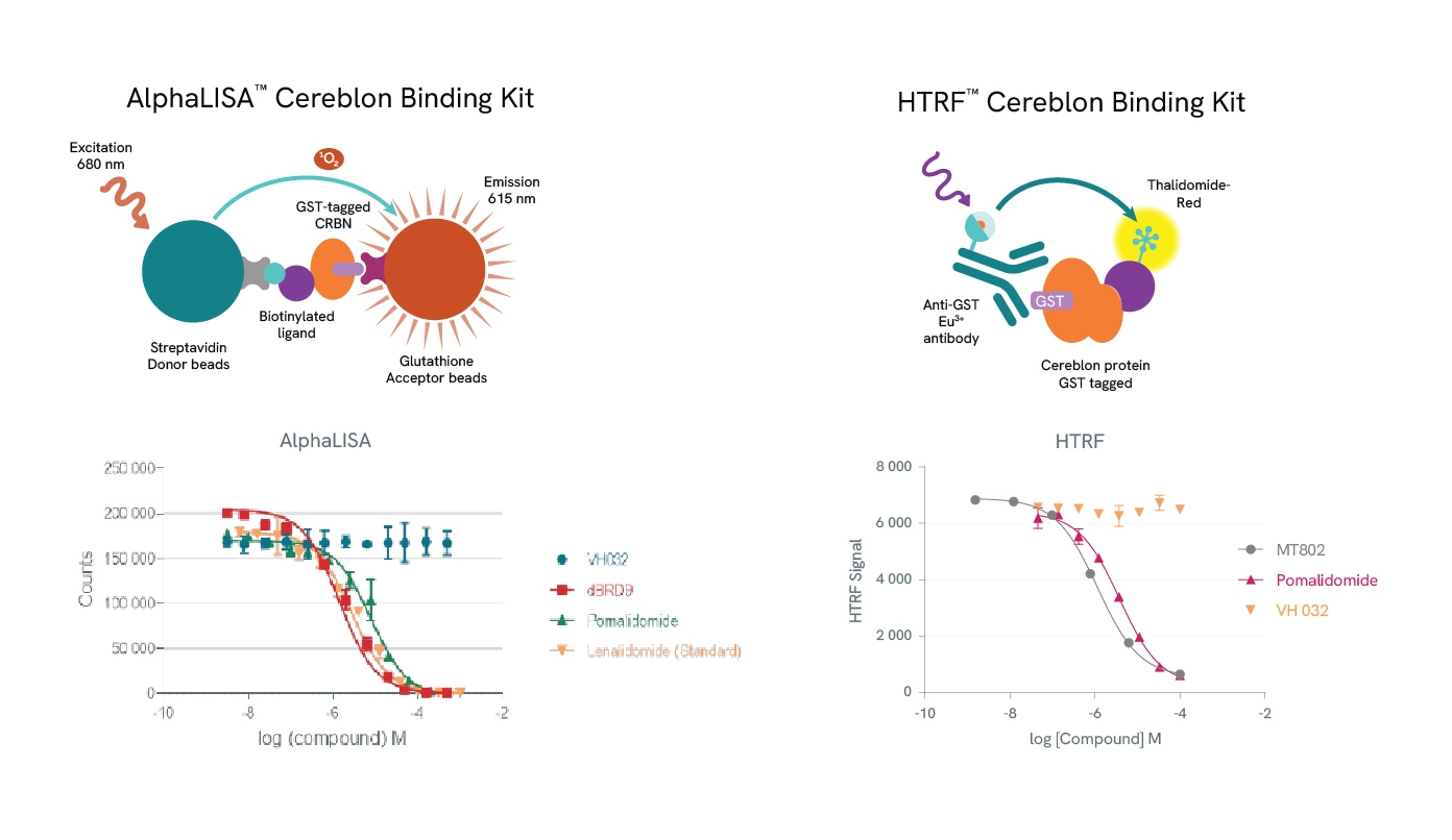
Top row: The AlphaLISA detection of Cereblon binding uses glutathione acceptor beads to capture GST-tagged Cereblon and Streptavidin-coated donor beads to capture the biotinylated ligand. Donor beads and acceptor beads come into proximity through ligand binding to Cereblon. In the HTRF assay Cereblon binding is detected using a specific GST antibody labeled with Europium Cryptate (donor) which binds to GST-tagged Cereblon and Thalidomide-Red labelled with XL665 (acceptor). Addition of a Cereblon binding compound leads to competitive displacement of the biotinylated ligand/Thalidomide red respectively, and a reduction of light emission inversely proportional to the compound’s concentration.

Bottom row: Binding of dBRD9, Pomalidomide or Lenalidomide to Cereblon reduces the signal of the AlphaLISA assay in a concentration-dependent fashion compared to the negative control VH032. Similarly for the HTRF assay, MT802 and Pomalidomide induce a reduction of FRET, whereas VH032 doesn’t.

Imagesection3-252x252

Is the ternary complex formed correctly?

The binding efficacy and affinity of the PROTAC molecule for the POI and the E3 ligase in the context of a binary complex cannot predict the stability of the ternary complex. Increasing evidence suggests that the degradation activity of a PROTAC is highly dependent on the stability of ternary complexes and the cooperativity of protein-protein interactions.

The binding efficacy and affinity of the PROTAC molecule for the POI and the E3 ligase in the context of a binary complex cannot predict the stability of the ternary complex. Increasing evidence suggests that the degradation activity of a PROTAC is highly dependent on the stability of ternary complexes and the cooperativity of protein-protein interactions.

Characterizing the ternary complex

Any pharmaceutical development workflow will need to scrutinize extensively the formation, population, stability, binding affinities, cooperativity, and kinetics of PROTAC ternary complexes. Alpha and HTRF protein-protein interaction assay kits are ideal companions for such studies. By titrating a PROTAC molecule with its target protein and E3 ligase, a bell-shaped curve can be produced by plotting Alpha and HTRF signals against PROTAC concentrations, as high PROTAC concentrations result in binary complexes (PROTAC-POI or PROTAC-E3 Ligase} which compete for the ternary complex (hook effect). The height of the curve is dependent on ternary complex concentration, enabling the classification of PROTACs by their ability to form ternary complexes. In addition to ternary complex formation, Alpha and HTRF assay kits can also be repurposed to determine the affinity of these complexes.
 

image-8-alphalisa-htrf-protein-protein-assays-800px


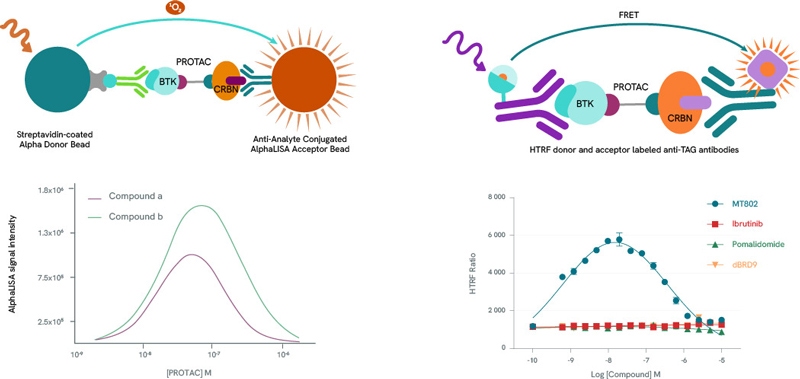
Left panel: Example bell curves constructed using an AlphaLISA protein-protein interaction assay. Compound a is more efficient than compound b in the formation of ternary complexes with BTK and Cereblon.

Right panel: Example bell-shaped curve obtained using an HTRF protein-protein interaction assay in the presence of BTK and Cereblon. Data from MT-802 demonstrate the formation of the ternary complex. As expected, the BTK inhibitor Ibrutinib, the Cereblon inhibitor Pomalidomide and the BRD9-specific PROTAC dBRD9 do not form ternary complexes.

Imagesection5-252x252

What does the future hold for Targeted Protein Degradation?

PROTAC technology is by no means the only player in the targeted protein technology field. A number of alternative methods have been developed that utilise both the UPS (TRAFTAC, molecular glues) as well as the ALS (LYTAC, AUTAC, ATTEC). Targeting the ALS in particular, offers some distinct advantages as it allows for the degradation of large proteins, macromolecular complexes or aggregates.

PROTAC technology is by no means the only player in the targeted protein technology field. A number of alternative methods have been developed that utilise both the UPS (TRAFTAC, molecular glues) as well as the ALS (LYTAC, AUTAC, ATTEC). Targeting the ALS in particular, offers some distinct advantages as it allows for the degradation of large proteins, macromolecular complexes or aggregates.

Molecular glues

In contrast to the PROTAC technology that relies on bifunctional degradation activation compounds (biDACs), molecular glues (or glue degraders) are monofunctional degradation activation compounds (monoDACs). These bind to either the E3 ligase or the target protein and chemically modify its surface, prompting protein-protein interactions that ultimately cause the E3 ligase to bind to the target protein, resulting in degradation. MonoDACs tend to be smaller molecules than biDACs, which could simplify optimization with respect to bioavailability and compliance with drug development guidelines. However, monoDACs’ reliance on single protein-protein interactions can weaken their ability to select the target of interest.

TRAFTAC

TRAnscription Factor TArgeting Chimeras (TRAFTACs) are heterobifunctional chimeric oligonucleotides. They consist of a double-stranded DNA moiety that recruits the transcription factor of interest (TOI), covalently linked to a Cas9 CRISPR-binding RNA. The latter binds to an ectopically expressed dCas9-HaloTag7 fusion protein. Combining the TRAFTAC with a PROTAC molecule specific for HaloTag7 and an E3 Ligase (VHL in the original study) brings into proximity the E3 Ligase and the DNA-bound TOI, resulting in the latter’s ubiquitination and proteasomal degradation.
 

image-12-traftac-and-molecular-glues-800px

ATTEC

An AuTophagosome TEthering Compound (ATTEC) is a bifunctional molecule capable of binding simultaneously to a target protein as well as the phagophore protein LC3. By doing so, the POI-ATTEC-LC3 complex is incorporated in a phagophore membrane, leading to its engulfment and proteolytic degradation once the phagophore merges with a lysosome to form an autophagosome.

AUTAC

The AUtophagy-TArgeted Chimera (AUTAC) consists of a specific binder for an intracellular target of interest and a guanine tag connected by a flexible linker. This induces S-guanylation of the target, and has been applied in the selective autophagic degradation of proteins as well as mitochondria. In the case of mitochondria, the guanine tag induced K63 poly-ubiquitination, which facilitated of mitophagy in a Parkin and PINK1-independent fashion.

LYTAC

Designed specifically to target cell membrane-bound and extracellular proteins, LYsosome-TArgeted Chimeras (LYTACs) make use of the lysosome degradation pathway by recruiting proteins to lysosome-shuttling receptors located at the cell surface. LYTACs consist of a POI-binding element linked to a moiety that allows transport into the cell via lysosome-shuttling receptors, where it will get degraded by the endosome/lysosome pathway. The first example of such a moiety consisted of a chemically synthesized glycopeptide ligand, an agonist of the cation-independent mannose-6- phosphate receptor (CI−M6PR). Another study focusing on Human hepatocytes (HCC) used the liver-specific asialoglycoprotein receptor (ASGPR) using a triantenerrary N-acetylgalactosamine (tri-GalNAc) motif as it displayed more efficient uptake than M6Pn-LYTAC owing to the higher surface expression of ASGPR in hepatocytes.
 

image-13-autophagy-lysosomal-system-800px


LYTAC, AUTAC and ATTEC mechanisms of action

As with PROTACs, our AlphaLISA and HTRF reagents can help research on the development of the other targeted degradation technologies, either in conjunction with the CETSA method, or standalone kits for transcription factors, kinases and protein-protein interactions.

bg-element-listing-page bg-element-listing-page-mobile
Products & Services
Resource Library
Filters
Expand All|Collapse All
Product Type
Application
Brand
Cellular or Signaling Pathway
Detection Modality
Host Species
Product Group
Target
Target Class
Target Species
Therapeutic Area
Sort by: Recommended
Recommended
Product Name A-Z
Product Name Z-A
Price - low to high
Price - high to low
1 - 25 of 223 Products and Services
AlphaLISA Surefire Ultra Total Protein

The AlphaLISA™ SureFire® Ultra™ Mouse Total RIPK1 assay is a sandwich immunoassay for quantitative detection of total RIPK1 in cellular lysates using Alpha Technology.

USD 694.00 - 46,060.00
AlphaLISA Surefire Ultra Total Protein

The AlphaLISA™ SureFire® Ultra™ Human Total Progesterone Receptor assay is a sandwich immunoassay for quantitative detection of total Progesterone Receptor in cellular lysates using Alpha Technology.

USD 694.00 - 46,060.00
AlphaLISA Surefire Ultra Total Protein

The AlphaLISA™ SureFire® Ultra™ Human Total p16 INK4A assay is a sandwich immunoassay for quantitative detection of total p16 INK4A in cellular lysates using Alpha Technology.

USD 694.00 - 46,060.00
AlphaLISA Surefire Ultra Total Protein

The AlphaLISA™ SureFire® Ultra™ Human Total PI3K p85 assay is a sandwich immunoassay for quantitative detection of total PI3K p85 in cellular lysates using Alpha Technology.

USD 694.00 - 46,060.00
AlphaLISA Surefire Ultra Total Protein

The AlphaLISA™ SureFire® Ultra™ Human and Mouse Total MEK2 assay is a sandwich immunoassay for quantitative detection of total MEK2 in cellular lysates using Alpha Technology.

USD 694.00 - 46,060.00
AlphaLISA Surefire Ultra Total Protein

The AlphaLISA™ SureFire® Ultra™ Human and Mouse Total NFκB2 p100 assay is a sandwich immunoassay for quantitative detection of total NFκB2 p100 in cellular lysates using Alpha Technology.

USD 694.00 - 46,060.00
AlphaLISA Surefire Ultra Total Protein

The AlphaLISA™ SureFire® Ultra™ Human and Mouse Total TRAF3 assay is a sandwich immunoassay for quantitative detection of total TRAF3 in cellular lysates using Alpha Technology.

USD 694.00 - 46,060.00
AlphaLISA Surefire Ultra Total Protein

The AlphaLISA™ SureFire® Ultra™ Human Total p21 CiP1 assay is a sandwich immunoassay for quantitative detection of total p21 CiP1 in cellular lysates using Alpha Technology.

USD 694.00 - 46,060.00
AlphaLISA Surefire Ultra Total Protein

The AlphaLISA™ SureFire® Ultra™ Human and Mouse Total NFκB2 p100/p52 assay is a sandwich immunoassay for quantitative detection of total NFκB2 p100/p52 in cellular lysates using Alpha Technology.

USD 694.00 - 46,060.00
AlphaLISA Surefire Ultra Total Protein

The AlphaLISA™ SureFire® Ultra™ Human and Mouse Total PRAS40 assay is a sandwich immunoassay for quantitative detection of total PRAS40 in cellular lysates using Alpha Technology.

USD 694.00 - 46,060.00
AlphaLISA Surefire Ultra Total Protein

The AlphaLISA™ SureFire® Ultra™ Human and Mouse Total BAD assay is a sandwich immunoassay for quantitative detection of total BAD in cellular lysates using Alpha Technology.

USD 694.00 - 46,060.00
AlphaLISA Surefire Ultra Total Protein

The AlphaLISA™ SureFire® Ultra™ Human Total SMARCA2 assay is a sandwich immunoassay for quantitative detection of total SMARCA2 in cellular lysates using Alpha technology.

USD 708.33 - 46,000.00
AlphaLISA Surefire Ultra Total Protein

The AlphaLISA™ SureFire® Ultra™ Human Total MYD88 assay is a sandwich immunoassay for quantitative detection of total MYD88 in cellular lysates using Alpha technology.

USD 708.33 - 46,000.00
AlphaLISA Surefire Ultra Total Protein

The AlphaLISA™ SureFire®  Ultra™ Human Total KRAS assay is a sandwich immunoassay for quantitative detection of total KRAS in cellular lysates using Alpha Technology.

USD 708.33 - 46,000.00
AlphaLISA Surefire Ultra Total Protein

The AlphaLISA™ SureFire®  Ultra™ Human and Mouse Total CDK8 assay is a sandwich immunoassay for quantitative detection of total CDK8 in cellular lysates using Alpha Technology.

USD 708.33 - 46,000.00
AlphaLISA Surefire Ultra Total Protein

The AlphaLISA™ SureFire® Ultra™ Human Total AIOLOS assay is a sandwich immunoassay for quantitative detection of total AIOLOS in cellular lysates using Alpha Technology.

USD 708.33 - 46,000.00
AlphaLISA Surefire Ultra Total Protein

The AlphaLISA™ SureFire®  Ultra™ Human and Mouse Total DJ1 assay is a sandwich immunoassay for quantitative detection of total DJ1 in cellular lysates using Alpha Technology.

USD 708.33 - 46,000.00
AlphaLISA Surefire Ultra Total Protein

The AlphaLISA™ SureFire®  Ultra™ Human Total Cyclin D2 assay is a sandwich immunoassay for quantitative detection of total Cyclin D2 in cellular lysates using Alpha Technology.

USD 708.33 - 46,000.00
AlphaLISA Surefire Ultra Total Protein

The AlphaLISA™ SureFire®  Ultra™ Human Total Cyclin D3 assay is a sandwich immunoassay for quantitative detection of total Cyclin D3 in cellular lysates using Alpha Technology.

USD 708.33 - 46,000.00
Picture of HTRF Human and Mouse LC3B-II Detection Kit

This HTRF kit enables the cell-based quantitative detection of LC3B-II (LC3-phosphatidylethanolamine conjugate).

Part Number: 64LC3B2PEG, 64LC3B2PEH
USD 2,271.53 - 13,214.42
Photo of KEAP1 Kit

The KEAP1 binding assay is designed to screen and characterize compounds that specifically bind to the E3 ligase, KEAP1.

Part Number: 64BDKEAP1PEG, 64BDKEAP1PEH
USD 1,304.58 - 15,640.42
Shipping box for Revvity reagent kits

The Total EGFR EX19DEL kit is designed to quantify the expression level of EGFR mutant DEL19 in cells

Part Number: 64EGFRD19PEG
USD 2,271.53
HTRF Human and Mouse Total Wee1 Detection Kit

The Total Wee1 kit is designed to monitor the expression level of cellular Wee1, a serine/threonine kinase.

Part Number: 64WEE1TPEG, 64WEE1TPEH
USD 2,271.53 - 13,214.42
Shipping box for Revvity reagent kits

The Total RBM39 kit is designed to quantify the expression level of RBM39 in cells

Part Number: 64RBM39TPEG, 64RBM39TPEH
USD 2,271.53 - 13,214.42
Shipping box for Revvity reagent kits

The Total PLK1 kit is designed to monitor the expression level of cellular human PLK1 and can be used as a normalization assay for the Phospho-human PLK1 Thr210 Detection Kit.

Part Number: 64PLKTPEG, 64PLKTPEH
USD 2,271.53 - 13,214.42
Filters
Expand All|Collapse All
Resource Type
Sort by: Resource Name A-Z
Resource Name A-Z
Resource Name Z-A
1 - 25 of 58 Resources
Application Note
Application Note
A guideline for HTRF cell-based phospho-protein data normalization

Get the best out of your phosphorylation assays Combining phospho and total protein assays enables better analysis. This Application Note provides valuable guidelines for efficiently analyzing and interpreting results from such assay combinations. Check out all the tips and examples in features! Features Introduction to phospho and total protein assay relevance Tips for handling and interpreting data Examples from actual experiments

Guide
Guide
Alpha Protein-Protein Interaction Quick Start Guide

Alpha has been used to study a wide variety of interactions, including protein:protein, protein:peptide, protein:DNA, protein:RNA, protein:carbohydrate, protein:small molecule, receptor:ligand, and nuclear receptor:ligand interactions. Both cell-based and biochemical interactions have been monitored, and applications such as phage display, ELISA, and EMSA (electrophoretic mobility shift assay) have been adapted to Alpha.

Brochure
Brochure
Alpha SureFire Ultra no-wash immunoassay catalog

Discover Alpha SureFire ®   Ultra ™ assays, the no-wash cellular kinase assays leveraging Revvity's exclusive bead-based technology and sandwich immunoassays for detecting phosphorylated proteins in cells. Offering a quantitative alternative to Western Blotting, Alpha SureFire assays are automation-friendly, easily miniaturized, and proficient in detecting both endogenous and recombinant proteins. Explore our comprehensive portfolio of SureFire Assays, designed to help you elevate and expedite your drug discovery journey.

Technical Note
Technical Note
AlphaLISA EZH2 Histone H3-Lysine 27 N-methyltransferase assay

Lysine 27 Dimethylation Detection with AlphaLISA Technology In this technical note discover how AlphaLISA immunodetection assay detects dimethylation of a biotinylated Histone H3 (21-44) peptide at lysine 27. It uses anti-di/mono-methyl-Histone H3 Lysine 27 (H3K27me2-1) as the detection antibody.

Technical Note
Technical Note
AlphaLISA G9a Histone H3-Lysine N-methyltransferase assay

Quantification of Histone H3 Lysine 9 Di-Methylation Using AlphaLISA Assay In this technical note discover how The AlphaLISA assay detects di-methylation of a biotinylated Histone H3 (1-21) peptide at lysine 9. In this assay, a biotinylated histone H3-derived peptide serves as the substrate, and the modified substrate is detected using Streptavidin (SA) Alpha Donor beads and AlphaLISA Acceptor beads conjugated to an antibody specific to the epigenetic mark of interest.

Technical Note
Technical Note
AlphaLISA SIRT1 Histone H3-Lysine 4 deacetylase assay

AlphaLISA Assay: A Tool for Measuring Histone H3 Deacetylation In this technical note, you will discover how the AlphaLISA immunodetection assay is used to determine the deacetylation level of a Histone H3 peptide (from the 1st to the 21st amino acid) that has been biotinylated and acetylated at the 4th lysine residue.

Technical Note
Technical Note
AlphaLISA SIRT1 p53 lysine 382 deacetylase assay

Measuring p53 Peptide Deacetylation: The AlphaLISA Assay Approach In this technical note, you will discover how the AlphaLISA immunoassay is used to detect the removal of acetyl groups from a biotin-labeled peptide derived from p53 (amino acids 368-393), specifically at the acetylated lysine residue number 382.

Guide
Guide
AlphaLISA SureFire Ultra assay optimization

This guide outlines further possible optimization of cellular and immunoassay parameters to ensure the best possible results are obtained.

Guide
Guide
AlphaLISA SureFire Ultra: the ultimate guide for successful experiments

The definitive guide for setting up a successful AlphaLISA SureFire Ultra assay Several biological processes are regulated by protein phosphorylation. It is, therefore, no surprise that the dysregulation of protein phosphorylation is implicated in a relatively large number of diseases. AlphaLISA SureFire Ultra assays provide a robust and reliable method for quantifying a targeted phosphorylation event in cell-based experiments. This guide contains tools and data helpful for you to perform your assays using AlphaLISA SureFire Ultra: A detailed description of the assay and its options A thorough investigation of assay conditions to obtain the optimal response from the chosen modulator and cell line A list of optimization steps to provide a sufficient assay window and produce the strongest results possible

Whitepaper
Whitepaper
An overview of atherosclerosis

Atherosclerosis pathogenesis, cellular actors, and pathways Atherosclerosis is a common condition in which arteries harden and become narrow due to a build-up of fatty material, usually cholesterol, and other substances such as calcium. This can lead to a range of serious health complications, including heart attack or stroke, making the disease an important contributing factor in death and morbidity in developed countries. Recent developments in our understanding of atherosclerosis from a molecular perspective include the discovery of new players in disease pathogenesis. Included in this white paper Atherosclerosis: step-by-step pathogenesis, therapeutic strategies, and recent developments Detailed descriptions and explanations, including a focus on pathways

eBook
eBook
Autophagy regulation eBook

Taking autophagy regulation research a step further Autophagy regulation is a key molecular process involved in recycling long-lived protein and organelles. Dysregulation of autophagy leads to different pathologies such as cancer, neurodegenerative and infectious diseases. This eBook features: Key facts about autophagy and mitophagy Infographics to apprehend the basics Cutting-edge knowledge

Guide
Guide
Benefit from an insight into the diversity of immune cells & signaling pathways

Get a useful overview of today’s immunity knowledge with this booklet Immunity is a collection of complex processes involving multiple strategies and specialized cell types. This booklet provides you with critical information regarding their roles, characteristic and signaling pathways as well as the collaborative behaviors that contribute to immunity. Featured in this guide: Review the fundamentals of immune cell types and mechanisms Learn from a cutting-edge research report Pathways and functional details on over 10 specialized immune cells

Technical Note
Technical Note
Best practices for analyzing brain samples with HTRF

Protocol for brain sample analysis with HTRF In this Technical Note, you will find tips we have complied to help ensure you are able to make the most of your brain sample. The Insider Tips you will find includes: Lysis step Determination of protein concentration Running of the optimized HTRF assay

Application Note
Application Note
CDK signaling in oncology: assess the effectiveness of a CDK9-targeting PROTAC molecule

CDK9 is an attractive target for cancer therapeutics due to its crucial role in transcription regulation, particularly of short-lived anti-apoptotic proteins such as MCL-1 and XIAP which are critical to the survival of cancer cells. One approach for specifically degrading CDK9 is using a proteolysis targeting chimera (PROTAC). In this application note, you will discover: The rapid and strong effect of CDK9 PROTAC molecule Thal-SNS-032, demonstrating specificity in CDK9 degradation How HTRF ® and AlphaLISA ® SureFire ® Ultra ™ are used to measure protein levels, with each tool showcasing the ability of PROTACs to rapidly degrade a targeted protein making it a unique tool in the fight against cancer

Application Note
Application Note
Characterizing chemokine receptor inhibitors with AlphaLISA SureFire Ultra, Alpha SureFire Ultra Multiplex and LANCE Ultra cAMP assays

The measurement of protein phosphorylation is a useful tool for measuring the modulation of receptor activation by both antibodies and small molecules. CCR7 and CXCR2 receptors, which are expressed in immune cells and are therapeutic targets for disorders like lupus erythematosus, adult leukemia, lymphomas, chronic obstructive pulmonary disease (COPD), and sepsis. AlphaLISA ™ SureFire ® Ultra ™ and Alpha SureFire ® Ultra ™ Multiplex assays are automation-friendly, applicable to both small and large-scale screens, and can assess phosphorylation status in complex matrices. The LANCE Ultra cAMP assay is measures cyclic AMP (cAMP) produced upon modulation of adenylyl cyclase activity by G-protein coupled receptors (GPCRs). This application note demonstrates how the SureFire Ultra and LANCE Ultra cAMP assays can be used for measuring inhibitors to CCR7 and CXCR2 cell surface receptors using a cellular model system where these receptors are overexpressed in CHO cells. The assays were optimized to measure receptor blockage and assayed receptor activity modulation by detecting ERK and AKT phosphorylation status and cAMP modulation. For more details, download the application note!

Technical Note
Technical Note
Detecting changes in phosphorylation events during the cell cycle

Lamin A/C, a crucial nuclear protein, undergoes phosphorylation at mitotic sites, controlled by the CDK1-Cyclin B1 complex, impacting cell cycle regulation, morphology, and fate. Dysregulation of phosphorylated Lamin A/C and associated proteins is linked to increased cell growth, genomic instability, and cell senescence in cancer, highlighting a promising target for therapeutic interventions. AlphaLISA ™ SureFire ® Ultra ™ technology provides a sensitive and efficient approach to monitor cell cycle dynamics and maintain cellular balance, even with low cell numbers. This technical note demonstrates its ability to detect changes in phosphorylated and total cell cycle regulatory proteins within the same cell lysate sample, offering a powerful tool for understanding complex cell biology and aiding drug discovery efforts. Learn more.

Application Note
Application Note
Detection of MAPK activation to evaluate the efficacy and potency of KRAS/SOS1 inhibitors by AlphaLISA and HTRF technologies

Evaluation of the therapeutic profile of anti-oncogene compounds in various cell lines with AlphaLISA™ and HTRF™ KRAS is a proto-oncogene known to be mutated in many cancer subtypes, inducing uncontrolled proliferation and cell metabolism changes. Like most small GTPases, KRAS will bind to GDP in its inactive form or to GTP in its active form. KRAS G12C is one of the most commonly found mutant forms in cancers, and leads to a permanently active state of KRAS. The upregulation of KRAS interaction with the exchange factor SOS1 leads to cancer phenotypes. Reducing KRAS activity and associated pathways could control the biological processes involved in cancer growth. Furthermore, it is well known that KRAS induces activation of mitogen-activated protein kinase (MAPK), thus playing a central role in human cancers. This application note provides a convincing demonstration of the reliability of the AlphaLISA and HTRF KRAS portfolios to evaluate compound in vitro therapeutic profiles in a cellular context: Determine the effects of KRAS and SOS1 inhibitors in different human cancer cell lines Discriminate the cellular action of KRAS-targeting compounds and evaluate their effectiveness in modulating KRAS downstream pathways.

Application Note
Application Note
Download your application note on nuclear receptor ligand identification with HTRF

Dive deeper into NASH investigation. Nuclear Receptors (NRs) regulate gene transcription by DNA binding. Interaction of NRs with an agonist triggers conformational changes and enables the recruitment of coactivators (CoA), leading to gene transcription. The activity of several NRs has been associated to NAFLD, bringing about the emergence of new therapeutic targets in NASH. Now, thanks to HTRF technology, you can monitor NR/CoA recruitment easily. With this application note you will: Benefit from HTRF technology in your research to identify potent NR ligands Discover the potential of HTRF PPi reagents to revolutionize your research Build robust and convenient NR/CoA recruitment assays for your research

Guide
Guide
Download your guide about neurodegenerative diseases

Emerging pathways to neuroinflammation and neurodegeneration Neurodegenerative diseases, such as amyotrophic lateral sclerosis, Parkinson's disease, or Alzheimer's disease, occur as a result of neuroinflammation and neurodegenerative progression. For the moment, these diseases are incurable. However, as research progresses, many similarities between these diseases have been found at a sub-cellular level. Review the fundamentals of the neuroinflammation process Learn from a cutting-edge research report Detailed insight into neurodegenerative diseases

Application Note
Application Note
Enhance PROTAC Drug Discovery with AlphaLISA and HTRF Kits

A Bruton Tyrosine Kinase (BTK) case study PROteolysis Targeting Chimeras (PROTACs) are one of the latest trending tools used in drug discovery. This note compiles experiments carried out to characterize PROTAC compounds using Bruton Tyrosine Kinase (BTK) as a model on Ramos cells. Both AlphaLISA and HTRF technologies were used in biochemical and cell-based contexts. Featured in this note: Assay principle and workflow Consistent results (data and graphs) Informative materials and methods, including references

Technical Note
Technical Note
Evaluating protein phosphorylation and cellular signaling cascades within the integrated stress response pathway

Protein phosphorylation, regulated by kinase and phosphatase cascades, plays a crucial role in cellular processes. Monitoring phosphorylation events in cellular models aids in studying compound activity and understanding mechanisms of action. In this technical note, we showcase the effectiveness of AlphaLISA ™ SureFire ® Ultra ™ assays in accurately measuring targeted phosphorylation events and total protein levels, with a focus on the eIF2α/ATF-4 pathway in cell-based experiments. Download the note to learn more!

Guide
Guide
Get your free HTRF™ guide

HTRF: unfamiliar territory? This technical brochure reviews the general principles of HTRF ™ and the associated Tag-lite ™ technology as well as discuss ways these technologies have been applied in drug discovery, biotherapeutics, and fundamental research in therapeutic areas such as Oncology, CNS, and Metabolic and Immune Disorders. Download a comprehensive guide to Homogeneous Time Resolved Fluorescence: HTRF features Benefits Targets, assays & applications

Technical Note
Technical Note
Guidelines for cell culture and lysis in different formats prior to HTRF detection

Adapt your cell culture plastic or glassware to your needs This Technical Note will provide you with guidelines on adapting the standard 96-well plate protocol for seeding and lysing cells to different culture formats such as T75 flasks, or 6-well, 12-well, and 24-well plates. Features Introduction to HTRF assays Recommended seeding, culture and lysis parameters for each cell culture container Demonstration case study of cells grown in various containers

Application Note
Application Note
Harness avidity without compromising on pharmacology

Characterize compounds in extra-low affinity interactions This Note groups protocols and data showing how a Biotin-Streptavidin system can be used to increase the apparent affinity of a protein-protein interaction and make it easy to characterize. No need to worry about the pharmacology of compounds tested this way: you will also see experiments demonstrating that this avidity-based approach does not impair compound characterization. Features: Apparent affinity of weak interactions enhanced with the Biot-SA system Evidence for pharmacological validation Kd determination using the avidity approach

Application Note
Application Note
High throughput platforms to characterize a bispecific antibody subjected to force degradation

Forced degradation studies are critical in therapeutic antibody research, helping to evaluate stability across the product lifecycle. Common degradation pathways include aggregation, deamidation, oxidation, and fragmentation, with thermal stress being a frequently studied factor. This application note explores the stability of a bispecific antibody targeting mouse CD200 and CD47 under thermal stress, using a high-throughput, no-wash HTRF™ assay to assess its binding activity. Read this application note to learn more. For research use only. Not for use in diagnostic procedures.