


Overview
Optical-based in vivo imaging of blood vessels and vascular leak is an emerging modality for studying changes that occur in a variety of different cancers and inflammatory states. A number of fluorescent imaging probes that circulate with the blood, but have no target selectivity, have been used to detect tumor leakiness as an indication of abnormal tumor vasculature. Inflammation is also characterized by distinct vascular changes, including vasodilation and increased vascular permeability, which are induced by the actions of various inflammatory mediators.
Revvity offers imaging probes that fluoresce in the near infrared (NIR) for detection of changes in vascular permeability.
Products for vascular imaging
| Probe | Probe type | Molecular weight/size | Excitation/emission wavelengths | Blood half-life | Tissue half-life |
|---|---|---|---|---|---|
| IVISenseTM Edema | BSA-targeted small molecule | 1540 g/mol | 675/692 nm | 1.5 h | 5 h |
| IVISense Vascular | PEGylated large scaffold | 250,000 g/mol | 680/700 nm | 7 h | 72 h |
| IVISense Vascular NP | Nanoparticle | 30-50 nm particles | 673/690 nm | 20 h | >100 h |
How to choose a probe for vascular imaging
NIR vascular probes can vary in size and physicochemical properties that affect their overall utility for particular imaging applications or affect the time window for optimal imaging. In studies, we compared three NIR fluorescent probes, IVISense Edema (a low molecular weight probe), IVISense Vascular (a high molecular weight probe), and IVISense Vascular NP (30-50 nm nanoparticles). Each probe differs significantly in pharmacokinetics, biodistribution, and tissue clearance rates, offering three slightly different imaging tools for cancer and inflammation research. All three probes can detect tumor vascular leak, with IVISense Vascular probe showing superior signal to background in orthotopic mouse breast cancer. In contrast, IVISense Edema probe shows superior imaging capability when applied to a mouse model of acute, inflammatory paw edema. IVISense Vascular NP, designed as a highly robust probe for intravital microscopy assessment of vessel morphometry, shows the ability to image both tumors and inflammation with long term accumulation at the imaging site (sometimes a benefit for long term studies). Images and quantification of time courses in animal models of cancer and inflammation provide guidance regarding optimal usage of these three vascular imaging probes.
Summary for four vascular applications
| IVISense Edema | IVISense Vascular | IVISense Vascular NP | |
|---|---|---|---|
| Tumor imaging | + | +++ | + |
| Tumor imaging time | 3 h | 24-48 h | 24-48 h |
| Tumor washout | 48 h | 144 h | >192 h |
| Response to antiangiogenic agents | nd | +++ | nd |
| Acute edema imaging | +++ | + | + |
| Tissue imaging time | 3 h | 24 h | 24 h |
| Tissue washout | 48 h | 144 h | >192 h |
| Arthritis imaging | nd | +++ | +++ |
| Tissue imaging time | nd | 24 h | 24 h |
| Tissue washout | nd | 144 h | >192 h |
| Intravital microscopy | ++ | +++ | +++ |
| Optimal imaging time | 5-15 min | 5-60 min | 5 min – 3 h |
Mouse model for imaging vascular disease
Imaging Tumors with Vascular Probes
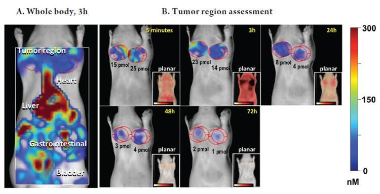
To determine the optimal imaging time points for the different vascular imaging probes, we used a syngeneic mouse tumor model, orthotopic implantation of 4T1 mouse breast adenocarcima at two sites (in each upper mammary fat pad).
Figure 1. IVISense Edema tumor imaging. Fluorescence molecular tomography and planar images show the patterns of fluorescence that occur in animals bearing tumor masses on the upper mammary fat pads. A) Whole body signal by fluorescence tomography imaging. B) Tomography imaging showing only tumor region fluorescence over time. Inset panels represent surface fluorescence detected by planar imaging.
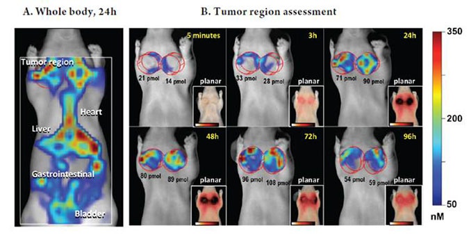
Figure 2. IVISense Vascular tumor imaging. Fluorescence tomography and planar images show the patterns of fluorescence that occur in animals bearing tumor masses on the upper mammary fat pads. A) Whole body signal using tomography imaging. B) Fluorescence tomography imaging showing only tumor region over time. Inset panels represent surface fluorescence detected by planar imaging.
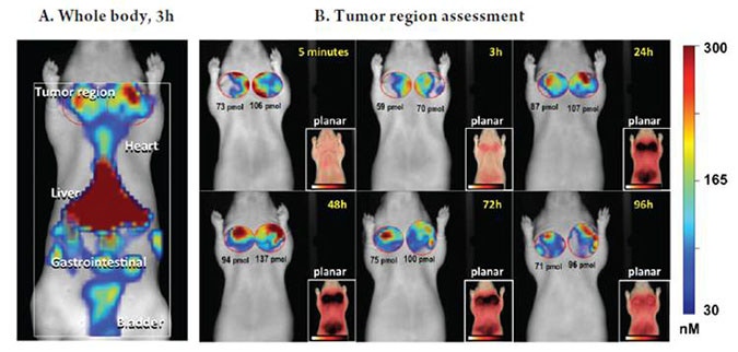
Figure 3. IVISense Vascular NP tumor imaging. Fluorescence tomography and planar images show the patterns of fluorescence that occur in animals bearing tumor masses on the upper mammary fat pads. A) Whole body signal using fluorescence tomography imaging. B) Tomoagraphy imaging showing only tumor region fluorescence over time. Inset panels represent surface fluorescence detected by planar imaging.
Imaging Acute Edema with vascular probes
We used a mouse model of acute neutrophil-driven paw edema induced by injection of carrageenan (CG) into the right hindpaws. This provided an experimental approach that permitted the comparison of fluorescence molecular tomography imaging and planar imaging in the analysis of vascular probe kinetics. IVISense Edema probe was injected at t = 2 h post carrageenan, whereas IVISense Vascular and IVISense Vascular NP were injected at t = 3 h, to accommodate the differences in blood pharmacokinetics and to permit imaging at the peak edema response (3 h). All three probes showed increased signal in carrageenan paws, although IVISense Vascular NP showed high background signal in control paws. IVISense Edema cleared quickly from tissue, making this probe ideal for imaging acute edema. Both IVISense Vascular and IVISense Vascular NP probes showed continued carrageenan paw accumulation at 24 h, a time at which there is less edema and more cellular inflammation.
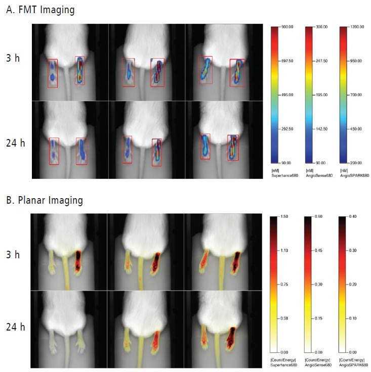
Figure 4. Imaging Acute Paw Edema Induced by Carrageenan Injection. Fluorescence molecular tomography and planar images show the patterns of fluorescence of three different vascular imaging probes that accumulate in paws of animals with inflammatory edema induced in the right footpad. A) Paw signal using fluorescence tomography. Numbers indicate quantitated pmol per paw. B) Planar imaging of the same mice shows the superficial fluorescence associated with the edema response.
For research use only. Not for use in diagnostic procedures