IVISense™Edema 680 (formerly Superhance) is a small molecule fluorescent in vivo blood pool imaging probe. IVISense Edema 680 enables imaging of circulation, blood vessels, vasculature, vascular leak, including that associated with early oncologic and opthalmologic lesions.
Products and catalog numbers
| Product | Catalog Number | Ex/Em wavelength (nm) | Molecular weight (g/mol) | Validated Experiments | Applications |
|---|---|---|---|---|---|
| IVISense Edema 680 | NEV10116 | 675/692 | ~ 1540 | In vivo | Vascular |
| Cancer/Tumor |
Using IVISense Edema 680 probe for in vivo studies
The generally recommended procedure for in vivo imaging with IVISense Edema 680 is administration via tail vein injection and imaging 0.5 – 24 hours post-injection.
- For Acute Edema, we recommend imaging 3 hours post injection.
- For IVM: 5-15 min post injection
| Product | Route of Injection | Mouse Dose (25 g) | Rat Dose (250 g) | Blood t 1/2 | Tissue t 1/2 | Optimal imaging time | Optimal Re-injection Time (complete clearance) | Route of Metabolism/ background tissue | FMT& IVIS settings |
|---|---|---|---|---|---|---|---|---|---|
| IVISense Edema 680 | IV | 4 nmol (150 uL) | 12-40 nmol | 1.5 h | 5 h | 3 h (1-3 h) | 96 h | Bladder | FMT 680/700 |
| IVIS 675/720 |
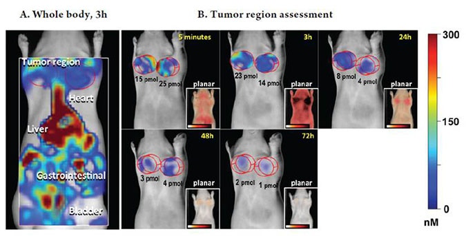
To determine the optimal imaging timepoint relative to probe injection, we imaged 4T1 tumor-bearing mice at different times post-IVISense Edema 680 injection (Figure 1). Whole body images at 3 h post-injection (Figure 1A) reveal signal throughout the body (including bladder, intestines, liver, and heart) as well as in the tumor periphery.

Figure 1. Imaging tumors. FMT and planar images show the patterns of fluorescence that occur in animals bearing tumor masses on the upper mammary fat pads. A) Whole body signal using fluorescence tomography imaging. B) Tomography imaging showing only tumor region fluorescence over time. Inset panels represent surface fluorescence detected by planar imaging.
Application notes and posters
Application note: Comparison of Revvity Vascular Pre-clinical Fluorescent Imaging Probes in Oncology and Inflammation Research
For research use only. Not for use in diagnostic procedures.
The information provided above is solely for informational and research purposes only. The information does not constitute medical advice and must not be used or interpreted as such. Consult a qualified veterinarian or researcher for specific guidance on injection techniques or other animal procedures. Revvity assumes no liability or responsibility for any injuries, losses, or damages resulting from the use or misuse of the provided information. The information is provided on an "as is" basis without warranties of any kind. Users are solely responsible for complying with all relevant laws, regulations, and institutional animal care and use committee (IACUC) guidelines in their use of the information provided.


