


Overview
Angiogenesis is the process through which new blood vessels form from pre-existing vessels. It is fundamental for tumor growth, progression and metastasis. Destroying tumor vasculature and/or inhibiting neovascularization have become well-accepted cancer treatment strategies.
Assessment of tumor size by caliper measurements is the most commonly used noninvasive metric of drug efficacy in preclinical research models. However, more invasive approaches are required to understand the underlying biology and the early changes associated with a particular cancer therapy. For example, the use of anti-angiogenic agents in combination with measuring tumor volume will most likely underestimate the effects of the treatment. This is because decreases in vascularity generally precede any effects on tumor size.
Fluorescence imaging approaches have been developed to better detect and monitor cancer. IVISense™ Vascular (formerly AngioSense) are fluorescent in vivo blood pool imaging probes. These probes are near-infrared labeled fluorescent macromolecules that remain localized in the vasculature for extended periods of time and enables imaging of blood vessels and angiogenesis.
Products and catalog numbers
| Product | Catalog Number | Ex/Em wavelength (nm) | Molecular weight (g/mol) | Validated Experiments | Applications |
|---|---|---|---|---|---|
| IVISense Vascular 680 | NEV10054EX | 670/690 | 70,000 | In vivo/Ex vivo Flow cytometry In vitro microscopy |
Vascularity in cancer Inflammation |
| IVISense Vascular 750 | NEV10011EX | 750/770 | 70,000 | In vivo/Ex vivo Flow cytometry In vitro microscopy |
Vascularity in cancer Inflammation |
Using IVISense Vascular for in vivo/ex vivo studies
The recommended procedure for in vivo imaging with IVISense Vascular 680 or IVISense Vascular 750 is administration via tail vein injection and imaging 24 hours post injection.
- Imaging in Oncology: IVISense Vascular 680 and IVISense Vascular 750 can be used to study angiogenesis, as a marker for blood vessel density, in animal tumor models.
- Imaging in Arthritis: IVISense Vascular 680 and IVISense Vascular 750 can be used to characterize vascular changes and therapeutic responses associated with animal models of arthritis.
- Instructions for setting up an in vivo mouse experiment and imaging on an IVIS imaging system with IVISense Vascular 750 fluroescent probe.
| Product | Route of Injection | Mouse Dose (25 g) | Rat Dose (250 g) | Blood t 1/2 | Tissue t 1/2 | Optimal imaging time | Optimal Re-injection Time (complete clearance) | Route of Metabolism/ background tissue | FMT and IVIS settings |
|---|---|---|---|---|---|---|---|---|---|
| IVISense Vascular 680 | IV | 2 nmol | 6-20 nmol | 7 h | 72 h | 24 h | 6-7 d | Low liver lung | FMT 680/700 IVIS 675/720 |
| IVISense Vascular 750 | IV | 2 nmol | 6-20 nmol | 16 h | 72 h | 24 h | 6-7 d | Low liver lung | FMT 750/770 IVIS 745/800 |
In Vivo/ex vivo imaging
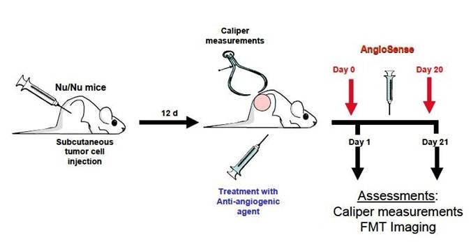
Figure 1: Optimal numbers of cultured Colo-205 tumor cells were implanted subcutaneously in the right flank of BALB/c nu/nu mice. After twelve days, tumors were measured using a conventional digital caliper, and mice were randomized (Day 0) into 2 groups of equivalent average starting tumor volumes. Compounds were formulated and injected intraperitoneally. Treatment with either vehicle or an anti-angiogenic agent (AAA) began on Day 0 and continued three times a week until Day 21. Mice were injected with IVISense Vascular 680 on Day 0 or 20 and imaged 24 hours later on an FMT in vivo imaging system. Tumor dimensions were measured by caliper throughout the study and tumor volumes were calculated using the formula volume (mm3) = (length x width3)
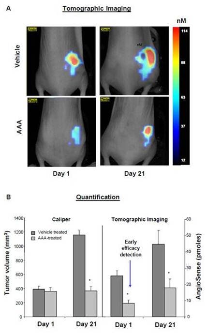
Figure 2: Imaging of mice treated as shown in Figure 1. A) Maximum projection slices were taken at the same color gating of a scan from representative untreated (top) and AAA-treated (bottom) mice. B) The total amount of fluorescence (in pmoles) was determined (right, tomographic imaging) and compared to tumor volume (left, caliper). The total fluorescence decreased 77% within 24 hours after the treatment. A quantifiable therapeutic response was detected, whereas no effect on tumor size could be shown using standard caliper assessment. By day 21, tumor volumes were statistically smaller in the treated animals and there was a statistically significant reduction in fluorescence. The background fluorescence showed no observable differences over time (data not shown).

Figure 3: 9L, HT-29, LLC and MS-1 tumor cells were implanted in the right flank of BALB/c Nu/Nu mice. When tumors reached an approximate volume of 60 mm3, mice were injected with IVISense Vascular 680 and imaged on the FMT 1 hour later. A) Immediately following imaging, mice were sacrificed, and tumors were excised. Tumor sections were stained with anti-CD31 antibody and the % of positive area was calculated using the Zeiss Axiovision software. Note the strong correlation between FMT signal and vascularization (r2 = 0.9983). B) Fluorescent signal within each tumor was captured by drawing a 3D region of interest (ROI) around the appropriate tumor area, and results were reported as peak concentration (in nM).
Application notes and posters
- Poster: A novel NIR dye for in vivo temporal tracking of labeled macrophages to sites of acute inflammation
- Poster: Non-Invasive Near-Infrared Fluorescence Quantitative Tomography (FMT ™) of the Effects of PDE4 Inhibitor Therapy in an LPS Murine Model of COPD In Vivo
- Poster: Quantitative Tomographic In Vivo Imaging of Syngeneic Breast Cancer Metastasis to the Lung and Therapeutic Response
FAQs
Q. Can I use IVISense Vascular probes in humans?
A. No, IVISense Vascular probes are intended for animal research use only, are not intended for use in diagnostic procedures and not intended for use in humans.
Q. I bought the AngioSense IVM previously, but I do not see it on your website.
A. We have discontinued the AngioSense IVM products.
Q. Can IVISense Vascular be used to detect vasculature? If so, how long should you wait to image mice to view vasculature?
A. Yes, our recommendation would be to image 30 min-1 hour after injection when the agent is high in circulation.
For research use only. Not for use in diagnostic procedures. The information provided above is solely for informational and research purposes only. The information does not constitute medical advice and must not be used or interpreted as such. Consult a qualified veterinarian or researcher for specific guidance or use information. Revvity assumes no liability or responsibility for any injuries, losses, or damages resulting from the use or misuse of the provided information, and Revvity assumes no liability for any outcomes resulting from the use or misuse of any recommendations. The information is provided on an "as is" basis without warranties of any kind. Users are responsible for determining the suitability of any recommendations for the user’s particular research. Any recommendations provided by Revvity should not be considered a substitute for a user’s own professional judgment. Users are solely responsible for complying with all relevant laws, regulations, and institutional animal care and use committee (IACUC) guidelines in their use of the information provided.