


Overview
Angiogenesis describes the formation of new blood vessels. Normal angiogenesis occurs during fetal development and wound healing. Deregulated angiogenesis is a key pathological event in cancer. In order to grow and metastasize, tumors need to continually acquire new blood supplies. Tumor angiogenesis describes the formation of new blood vessels that grow into the tumor, giving it nutrients and oxygen to assist its growth.
Anti-angiogenic agents have emerged as viable anti-cancer therapies, either alone or in combination with standard chemotherapy. Several classes of natural and synthetic agents target vascular endothelial cells, inhibiting the growth and development of new vessels. Anti-angiogenic agents include growth factor receptors, tyrosine kinase receptors, G-protein–coupled receptors for angiogenesis modulating proteins, integrins, matrix metalloproteinases inhibitors and adnectins.
In vitro and ex vivo models of angiogenesis can be used to provide insight on the molecular and cellular regulations involved in tumor vascularization and have enabled target screening and drug discovery. In vivo models can also be used to provide biological insights to angiogenic and antiangiogenic processes, as well as to evaluate drug efficacy and monitor tumor response in cancer therapies.
Products for imaging angiogenesis
We have validated the following IVISense™ fluorescent probes for imaging neovasculature associated with angiogenesis.
| Probe/Dye | Probe/dye type | Probe/Dye Mechanism | Available Wavelengths and optimal in vivo imaging time (post-injection) | Route of metabolism/ background tissue(s) | Validated imaging methods |
|---|---|---|---|---|---|
| IVISense Integrin Receptor | Targeted; fluorescent | Binds to α5β3 integrin. This integrin is upregulated in activated endothelial cells during angiogenesis. | 645 nm (6-24 hours) 680 nm (3-48 hours) 750 nm (24 hours) |
Bladder/kidneys (645 nm) Kidneys/intestine (680 nm) Kidneys (750 nm) |
In vivo/ex vivo; in vitro microscopy |
| IVISense Hypoxia CA IX | Targeted; fluorescent | Binds to carbonic anhydrase IX which is expressed in hypoxic tissue, a result of abherrant vasculature. | 680 nm (24 hours) | Kidneys | In vivo/ex vivo; flow cytometry; in vitro microscopy |
| IVISense Tomato Lectin | Targeted; fluorescent | Binds to tomato lectin, a well-known tool for studying tumor angiogenesis and measuring microvessel density. This probe provides direct labeling of the vascular endothelial cells. | 680 nm (6 h) | Overall vascular background | In vivo/ex vivo; flow cytometry; in vitro microscopy |
| IVISense Vascular | Vascular; fluorescent | A high molecular weight probe with a moderate blood half-life (7 hours), used to assess vascular leak, with ideal imaging times of 24 hours post-injection. | 680 nm (24 hours) 750 nm (24 hours) |
Low liver lung | In vivo/ex vivo; flow cytometry; in vitro microscopy |
| IVISense Vascular NP | Vascular; fluorescen | Pegylated fluorescent nanoparticles that remain localized in the vasculature for extended periods of time and enable imaging of blood vessels and angiogenesis | 680 nm (0 - 4 hours) | Long term tissue accumulation | In vivo |
| IVISense Edema | Vascular; fluorescent | Blood pooling probe | 680 nm (0.5 - 24 hours) | Bladder | In vivo |
IVISbrite™ Bioluminescent Tumor Cell Lines can also be implanted into mice to generate tumors and then can be used in combination with the fluorescent probes above to study angiogenesis and the effect of anti-angiogenic treatment on tumor volume.
Choosing the right probe
When choosing a probe for angiogenesis studies, several factors should be taken into consideration:
- Fluorescent vs. bioluminscent probes: Be sure to choose the proper probe for your instrumentation. Some imagers can measure both bioluminescence and fluorescence, while others can only measure one or the other.
- Dye excitation/emission wavelength: Some fluorescent probes are available with excitation wavelengths that range from 645 nm to 800 nm. Be sure to pick a wavelength that is appropriate for both your instrument and application. Most microscopes filters are not suitable for wavelengths above 680 nm. However deep tissue imaging typically has less background fluorescence and works better with longer wavelengths (750 nm- 800 nm).
- Probe clearance time is typically faster for activatable probes, which allows for re-injection after shorter time periods.
- Probe specificity: For targeted and activatable probes, it is important to remember that the protein or enzyme that the probe is targeting needs to be present and well expressed in your mouse model.
- In vivo distribution: Some probes might accumulate in organs at timepoints which could interfere with your study. Be sure to check the biodistribution profile of each probe of interest.
- Type of imaging required for your study: Vascular probes for example, while useful in assessing vascularity changes in the mouse, do not make good probes for in vitro cell imaging or tissue analysis. If these types of analyses are desired, targeted or activatable probes might be a better choice for you.
Mouse model for imaging angiogenesis
Cell lines
Rat gliosarcoma 9L, human colorectal adenocarcinoma HT-29, mouse Lewis lung carcinoma LLC, primary islet endothelial MS-1, mouse breast carcinoma 4T-1, and human colorectal adenocarcinoma Colo-205 lines were purchased from ATCC and cultured in recommended media supplemented with 10% FBS at 37 ºC.
Pharmacokinetics
Mice were injected with 2 nmoles of IVISense Vascular 680. At different times, blood was collected in heparinized tubes and the fluroescence measured in a Gemini fluorescence microplate reader (Molecular Devices).
Mouse models
BALB/c nude mice were subcutaneously implanted with optimal numbers of cultured tumor cells, and mouse tumors were imaged 1-24 hrs post-probe injection. For treatment studies, mice with established tumors were later randomized into 2 groups (Vehicle and CT-322) of equivalent average starting tumor volumes. Compounds were formulated and injected intraperitoneally. Tumor volumes were measured using the formula volume (mm3) = (length x width3)/2. APC (min) mice were purchased from Jackson Laboratories and placed onto a high fat diet. When fed a high fat diet, these mice develop spontaneous colon polyps around 3 months of age.
Figure 1. Experimental protocol for testing anti-angiogenic adnectin CT-322 (Adnexus, a Bristol Myers Squibb R&D company) in a tumor xenograft model using IVISense Vascular (formerly AngioSense®) for imaging.
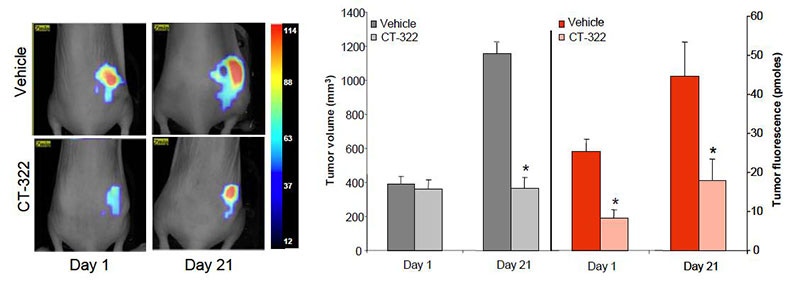
Figure 2. Imaging and quantification of the effects of the anti-angiogenic adnectin CT-322 on tumor vascular fluorescence using IVISense Vascular.
Application notes and posters
- Poster: Near-Infrared Fluorescence Imaging and Quantification of Anti-Angiogenic Therapy using an α5β3 Integrin-Targeted Probe
- Poster: In Vivo Quantification of Integrin-Targeted and Protease-Activated Imaging Probes in Response to Anti-Angiogenic Therapy Using Quantitative Fluorescence Tomography
For research use only. Not for use in diagnostic procedures. The information provided above is for informational and research purposes only. Revvity assumes no liability for any injuries, losses, damages, or outcomes arising from the use or misuse of this information or its recommendations. This information is provided "as is" without warranties. Users are solely responsible for determining the suitability of recommendations for their research and should not substitute them for their own professional judgment.