


Overview
The protein tomato (L. esculentum) lectin is widely used for vascular labeling because the protein has high binding affinity for glycoprotein N-acetylglucosamines on the surface of vascular endothelial cells. IVISense™ Tomato Lectin 680 (formerly TLectinSense 680) is a near-infrared fluorescent imaging probe which is comprised of the tomato lectin protein and a near-infrared fluorophore. IVISense Tomato Lectin 680 targets the vasculature and enables imaging of blood vessels and angiogenesis. It was designed and optimized for use in living animals. It can be used in assessing vascularity in vivo and in real time, without termination of mice, excision and processing of the tissue. It can also be used for improving the efficacy, early detection and monitoring of anti-angiogenic therapies in animal models.
Tumors induce significant blood vessel development in order to support their aggressive growth and progression. The extensive and disordered nature of tumor vasculature impairs drug delivery and efficacy. Therefore, destroying the tumor vasculature and/or inhibiting neo-vascularization have become well-accepted and proven cancer treatment strategies. IVISense Tomato Lectin 680 targets the surface of vascular endothelial cells and can be used for quantitation of vascular burden across different tumor cell lines.
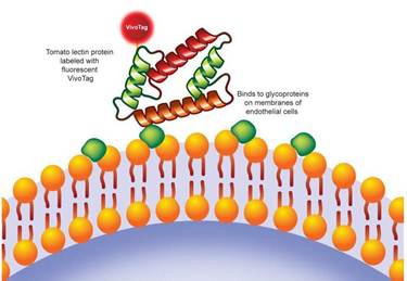
Figure 1: IVISense Tomato Lectin 680 mechanism of action. A near infrared IVISense 680 NHS Fluorescent Dye (formerly VivoTag 680 XL) was conjugated to the tomato lectin protein to produce the imaging probe.
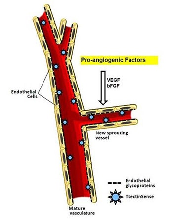
Figure 2: Tomato lectin is a recognized approach to labeling the vascular endothelium, intestinal epithelium and microglia. The mechanism for targeting vascular endothelium with tomato lectin is an established gold standard.
Products and catalog numbers
| Product | Catalog Number | Ex/Em wavelength (nm) | Molecular weight (g/mol) | Validated Experiments | Applications |
|---|---|---|---|---|---|
| IVISense Tomato Lectin 680 | NEV10060 | 670/690 | 72,000 | In vivo/ex vivo | Angiogenesis |
| Flow cytometry | Inflammation | ||||
| In vitro microscopy | Vascular Disease |
IVISense Tomato Lectin 680 probe for in vivo/ex vivo studies
The recommended procedure for in vivo imaging with IVISense Tomato Lectin 680 is intravenous administration and imaging 6 hours post injection.
Imaging in matrigel plugs and tumor models: IVISense Tomato Lectin 680 can be used to study angiogenesis and blood vessel density, in matrigel plugs and animal tumor models.
View instructions on setting up an in vivo imaging mouse experiment with IVISense Tomato Lectin 680 fluorescent probe.
| Route of Injection | Mouse Dose (25 g) | Rat Dose (250 g) | Blood t 1/2 | Tissue t 1/2 | Optimal imaging time | Optimal Re-injection Time (complete clearance) | Route of Metabolism/ background tissue | FMT and IVIS settings |
|---|---|---|---|---|---|---|---|---|
| IV | 2 nmol | 6 nmol | 2 h | >48 h | 6 h | 4-6 d | Overall vascular background | FMT 680/700 |
| IVIS 675/720 |
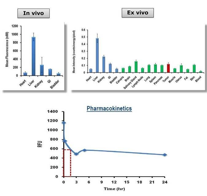
Figure 3: A) LLC tumor-bearing mice were injected with IVISense Tomato Lectin 680, imaged tomographically at 6 hours and fluorescence was quantified in 5 different organs. Tissues were then collected, and fluorescence assessed by planar imaging (ex vivo). Mean counts/energy for each tissue were determined as a measure of tissue brightness. B) The plasma pharmacokinetic profile was assessed by injecting CD1 mice (3/time point), collecting plasma at multiple times post-injection, and measuring plasma fluorescence.
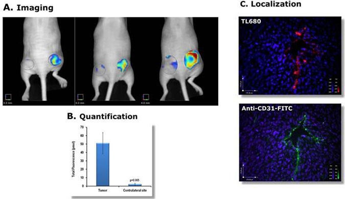
Figure 4: A) Lewis Lung Carcinoma cells were injected into the right flanks of Nu/Nu female mice and grown for 10 days. Mice were imaged tomographically (FMT 2500LX) 6 hours after intravenous IVISense Tomato Lectin 680 injection (2 nmoles; three representative mice are shown). B) Quantification of the tumor and contralateral site signal (pmoles) and C) Shows localization of the fluorescent signal in a frozen tumor section (Blue: DAPI nuclear staining, Red: IVISense Tomato Lectin 680, Green: anti-CD31).
Ex Vivo imaging
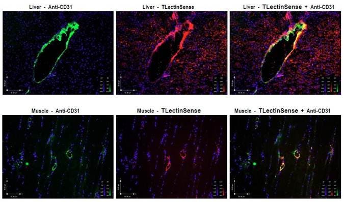
Figure 5: Labels vasculature in normal tissues. In vivo administration of IVISense Tomato Lectin 680 (formerly TLectinSense 680) also shows labeling of the liver and muscle vasculature, as confirmed by ex vivo co-localization with Anti-CD31-FITC.
Ex Vivo imaging
IVISense Tomato Lectin 680 enables imaging of endothelial glycoproteins in endothelial cells such as HUVEC.
Flow cytometry and in vitro microscopy
We have validated IVISense Tomato Lectin 680 for use with fluorescence microscopes and flow cytometers. Here is a brief protocol with a recommended concentration of probe to use:
- Culture cells in standard TC plate or chamber slide.
- Incubate cells with 0.25 µM IVISense Tomato Lectin 680 for 30 min at 37 °C.
- Wash 2x with PBS. For flow cytometry, detach and resuspend cells in PBS.
- Flow cytometry filter settings: 712/21 Fluorescence microscopy filter: Cy5.5
In Vitro Imaging:
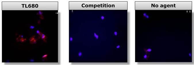
Figure 6: Human umbilical vein endothelial cells (HUVEC) were incubated with IVISense Tomato Lectin 680 in the absence (TL680) or presence (Competition) of 50X unlabeled lectin and visualized by fluorescence microscopy. DAPI nuclear staining is in blue and IVISense Tomato Lectin 680 signal is shown in red.
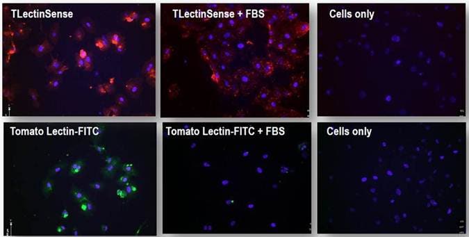
Figure 7: IVISense Tomato Lectin 680 (formerly TLectinSense 680) binds to human umbilical vein endothelial cells (HUVEC). The effect of plasma on binding was tested by incubating the probes/agents (1 mM or 20 mg/ml) in either PBS or 100% FBS. As a control, commercially available Lectin-FITC (20 mg/ml) was used. In blue: DAPI nuclear stain, red: IVISense Tomato Lectin 680, Green: Lectin-FITC. Binding of IVISense Tomato Lectin 680 to HUVEC is not inhibited by plasma (FBS), whereas binding of commercial lectin-FITC is inhibited.
Application notes and posters
- Poster: Targeted in vivo imaging of tumor vasculature using a near infra-red labeled tomato lectin probe
For research use only. Not for use in diagnostic procedures.