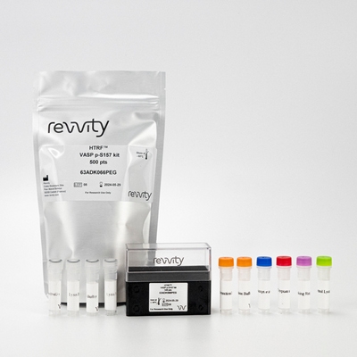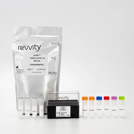

HTRF Human & Mouse Phospho-VASP (Ser157) Detection Kit, 500 Assay Points


HTRF Human & Mouse Phospho-VASP (Ser157) Detection Kit, 500 Assay Points


The phospho-VASP (Ser157) enables the cell-based quantitative detection of VASP phosphorylated on Ser 157 for monitoring PKA and PKG activation.
| Feature | Specification |
|---|---|
| Application | Cell Signaling |
| Sample Volume | 16 µL |
The phospho-VASP (Ser157) enables the cell-based quantitative detection of VASP phosphorylated on Ser 157 for monitoring PKA and PKG activation.


HTRF Human & Mouse Phospho-VASP (Ser157) Detection Kit, 500 Assay Points


HTRF Human & Mouse Phospho-VASP (Ser157) Detection Kit, 500 Assay Points


Product information
Overview
Phospho-VASP (Ser157) cellular assay is ideal for monitoring PKA activation, which phosphorylates VASP on Serine 157. VASP is involved in cell motility, migration, and adhesion. This makes phospho-VASP assaying a valuable tool in cardiovascular, oncology, and inflammation research.
Specifications
| Application |
Cell Signaling
|
|---|---|
| Brand |
HTRF
|
| Detection Modality |
HTRF
|
| Lysis Buffer Compatibility |
Lysis Buffer 1
Lysis Buffer 2
Lysis Buffer 3
Lysis Buffer 4
Lysis Buffer 5
|
| Molecular Modification |
Phosphorylation
|
| Product Group |
Kit
|
| Sample Volume |
16 µL
|
| Shipping Conditions |
Shipped in Dry Ice
|
| Target Class |
Phosphoproteins
|
| Target Species |
Human
Mouse
|
| Technology |
TR-FRET
|
| Therapeutic Area |
Cardiovascular
Metabolism/Diabetes
|
| Unit Size |
500 Assay Points
|
Video gallery

HTRF Human & Mouse Phospho-VASP (Ser157) Detection Kit, 500 Assay Points

HTRF Human & Mouse Phospho-VASP (Ser157) Detection Kit, 500 Assay Points

Citations
How it works
Phospho-VASP (Ser157) assay principle
The Phospho-VASP (Ser157) assay measures VASP when phosphorylated at Ser157. Contrary to Western Blot, the assay is entirely plate-based and does not require gels, electrophoresis or transfer. The Phospho-VASP (Ser157) assay uses 2 labeled antibodies: one with a donor fluorophore, the other one with an acceptor. The first antibody is selected for its specific binding to the phosphorylated motif on the protein, the second for its ability to recognize the protein independent of its phosphorylation state. Protein phosphorylation enables an immune-complex formation involving both labeled antibodies and which brings the donor fluorophore into close proximity to the acceptor, thereby generating a FRET signal. Its intensity is directly proportional to the concentration of phosphorylated protein present in the sample, and provides a means of assessing the proteins phosphorylation state under a no-wash assay format.

Phospho-VASP (Ser157) 2-plate assay protocol
The 2 plate protocol involves culturing cells in a 96-well plate before lysis then transferring lysates to a 384-well low volume detection plate before adding phospho-VASP (Ser157) HTRF detection reagents. This protocol enables the cells' viability and confluence to be monitored.

Phospho-VASP (Ser157) 1-plate assay protocol
Detection of Phosphorylated VASP (Ser157) with HTRF reagents can be performed in a single plate used for culturing, stimulation and lysis. No washing steps are required. This HTS designed protocol enables miniaturization while maintaining robust HTRF quality.

Assay validation
Validation on A431cells treated with forskolin
Human A431 cells were plated at 100,000 cells/well in a 96 well plate, and incubated for 24h at 37°C, 5% CO2. After treatment for 30 min with increasing concentrations of Forskolin, the medium was removed and the cells were lysed with 60 µL of lysis buffer for 30min at RT under gentle shaking. 16 µL of lysate were transferred into a 384-well low volume white microplate and 4 µL of the HTRF phospho-VASP (Ser157), phosphor-VASP (S239) or total VASP detection reagents were added. The HTRF signal was recorded after an overnight incubation.

Validation on NIH3T3 cells treated with forskolin
Mouse NIH3T3 cells were plated at 100,000 cells/well in a 96 well plate, and incubated for 24h at 37°C, 5% CO2. After treatment for 30 min with increasing concentrations of Forskolin, the medium was removed and the cells were lysed with 60 µL of lysis buffer for 30 min at RT under gentle shaking. 16 µL of lysate were transferred into a 384-well low volume white microplate and 4 µL of the HTRF phospho-VASP (Ser157), phosphor-VASP (S239) or total VASP detection reagents were added. The HTRF signal was recorded after an overnight incubation.

Simplified pathway
Function and regulation of VASP
Vasodilator-stimulated phosphoprotein (VASP) is an actin-associated protein and a member of the Ena-VASP family. VASP stimulates actin filament elongation, and is involved in cytoskeleton remodeling and cell polarity. VASP proteins are involved in axon guidance, platelet activation and cell migration. In platelets, VASP is a major substrate for cAMP-dependent protein kinase A (PKA) and cGMP-dependent protein kinase G (PKG). Whereas the preferred site for PKA is Ser-157, the preferred site for PKG is Ser-239. Phosphorylation modulates F-actin binding, actin filament elongation and platelet activation. VASP phosphorylation is often used to monitor the effect of drugs that reduce platelet reactivity in cardiovascular diseases.

Resources
Are you looking for resources, click on the resource type to explore further.
Atherosclerosis pathogenesis, cellular actors, and pathways
Atherosclerosis is a common condition in which arteries harden and...


How can we help you?
We are here to answer your questions.


