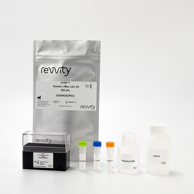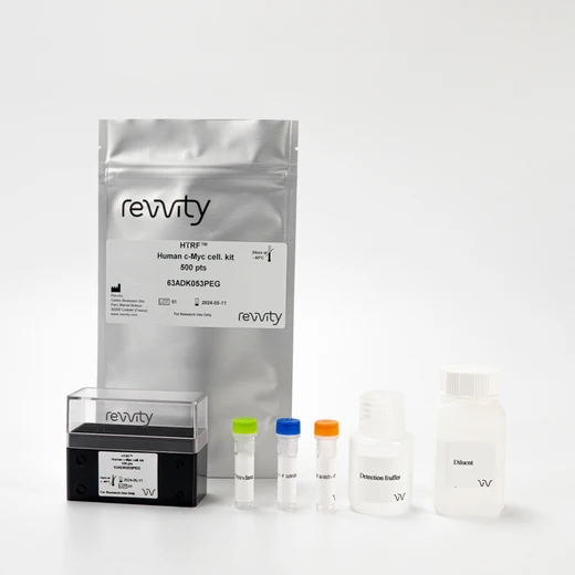

HTRF Human c-Myc Detection Kit, 500 Assay Points


 View All
HTRF Human c-Myc Detection Kit, 500 Assay Points


The Human c-Myc Cell-based kit is designed for the rapid detection of human c-Myc proto-oncogene in cells.
For research use only. Not for use in diagnostic procedures. All products to be used in accordance with applicable laws and regulations including without limitation, consumption and disposal requirements under European REACH regulations (EC 1907/2006).
| Feature | Specification |
|---|---|
| Application | Protein-Protein Interaction |
| Sample Volume | 10 µL |
The Human c-Myc Cell-based kit is designed for the rapid detection of human c-Myc proto-oncogene in cells.
For research use only. Not for use in diagnostic procedures. All products to be used in accordance with applicable laws and regulations including without limitation, consumption and disposal requirements under European REACH regulations (EC 1907/2006).


HTRF Human c-Myc Detection Kit, 500 Assay Points


HTRF Human c-Myc Detection Kit, 500 Assay Points


Product information
Overview
Human c-Myc is a multifunctional transcription factor protein that plays a role in cell cycle progression, apoptosis, and cellular transformation. c-Myc also functions to regulate global chromatin structure by regulating histone acetylation. The kit is intended for the specific and fast quantification of c-Myc proto-oncogene in cells.
Specifications
| Application |
Protein-Protein Interaction
|
|---|---|
| Brand |
HTRF
|
| Detection Modality |
HTRF
|
| Product Group |
Kit
|
| Sample Volume |
10 µL
|
| Shipping Conditions |
Shipped in Dry Ice
|
| Target Class |
Binding Assay
|
| Target Species |
Human
|
| Technology |
TR-FRET
|
| Therapeutic Area |
Oncology & Inflammation
|
| Unit Size |
500 Assay Points
|
Video gallery

HTRF Human c-Myc Detection Kit, 500 Assay Points

HTRF Human c-Myc Detection Kit, 500 Assay Points

How it works
Assay principle
The Human c-Myc is a sandwich immunoassay involving two specific anti-human c-Myc antibodies, respectively labelled with Europium Cryptate (donor) and d2 (acceptor). The intensity of the signal is proportional to the concentration of c-Myc present in the sample. The protocol is optimized for a 384-well plate format, but can easily be further miniaturized or upscaled. Only low sample volumes are needed. The detection reagents may be pre-mixed and added in a single dispensing step for direct detection. No washing is needed at any step.

Assay protocol
Two-plate assay protocol The human c-Myc assay protocol is described here. Cells are plated, stimulated, and lysed in the same 96-well culture plate. Lysates are then transferred to the assay plate for the detection of human c-Myc by HTRF reagents. This protocol enables the cells' viability and confluence to be monitored. The antibodies labelled with HTRF fluorophores may be pre-mixed and added in a single dispensing step to further streamline the assay procedure. The assay detection can be run in 96- to 384-well plates by simply resizing each addition volume proportionally.

Assay validation
HTRF assay compared to western blot
HeLa cells were grown in a T175 flask at 37°C - 5% CO2 for 48 h. After medium removal, the cells were lysed with 3 mL of supplemented lysis buffer for 45 min at RT. Serial dilutions of the cell lysate were performed in the supplemented lysis buffer and 10 µL of each dilution were dispensed and analyzed side-by-side by Western-blot and by HTRF. By using HTRF c-Myc only 1,000 cells are sufficient for minimal signal detection while 4,000 cells are needed for a Western Blot signal. The HTRF c-Myc assay is at least 4-fold more sensitive than the Western Blot.

c-Myc detection in several human cell lines
100,000 human MCF7, A549, HeLa, HTC116 and HEK293T cells were plated in 96-well plates in cell culture medium and incubated for 24h at 37°C - 5% CO2. Cell culture medium was then removed and cells were lysed with 50 µL of lysis buffer for 30 min at RT under gentle shaking. 10 µL of lysate was transferred into a 384-well sv white microplate, and 10 µL of the HTRF c-Myc detection reagents were added. The HTRF signal was recorded after an overnight incubation time.

Bromodomain interaction inhibitor effect
40,000 HeLa cells were plated in 96 well plate in cell culture medium and incubated for 24h at 37°C - 5% CO2. After incubating with increasing concentrations of JQ1 for 24 hours, the medium was removed and cells were lysed with 50 µL of lysis buffer for 45 min at RT under gentle shaking. 10 µL of lysate were transferred into a 384-well sv white microplate and 10 µL of the HTRF c-Myc detection reagents were added. The HTRF signal was recorded after an overnight incubation.

Histone deacetylase (HDAC) inhibitor effect
40,000 HeLa cells were plated in 96-well plates in cell culture medium, and incubated for 24h at 37°C - 5% CO2. After incubating with increasing concentrations of TSA for 24 hours, the medium was removed and cells were lysed with 50 µL of lysis buffer for 45 min at RT under gentle shaking. 10 µL of lysate were transferred into a 384-well sv white microplate and 10 µL of the HTRF c-Myc detection reagents were added. The HTRF signal was recorded after an overnight incubation.

Cell growth inhibitors effect
10,000 HeLa or A549 cells were plated in 96-well plates in cell culture medium and incubated for 24h at 37°C - 5% CO2. After incubating with increasing concentrations of inhibitors (U0126, Methotrexate and Gemcitabine) for 72 hours, the medium was removed and cells were lysed with 50 µL of lysis buffer for 45 min at RT under gentle shaking. 10 µL of lysate were transferred into a 384-well sv white microplate and 10 µL of the HTRF c-Myc detection reagents were added. The HTRF signal was recorded after an overnight incubation

Resources
Are you looking for resources, click on the resource type to explore further.
Discover the versatility and precision of Homogeneous Time-Resolved Fluorescence (HTRF) technology. Our HTRF portfolio offers a...
This guide provides you an overview of HTRF applications in several therapeutic areas.
An in-depth review of molecular and cellular pathways
The maintenance of proteostasis, the biological mechanisms that control the...


How can we help you?
We are here to answer your questions.


