


US
Search Results
Revvity Sites Globally
Select your location.
*e-commerce not available for this region.

HTRF Human and Mouse Phospho-β-Catenin (Thr41/Ser37/Ser33) Detection kit, 10,000 Assay Points


B-catenin P-t41/s33/37 Kit- 50,000 Tests
For research use only. Not for use in diagnostic procedures. All products to be used in accordance with applicable laws and regulations including without limitation, consumption & disposal requirements under European REACH regulations (EC 1907/2006).
Product Variants
Product information
Overview
The Phospho ß-Catenin (Thr41/Ser37/Ser33) assay kit offers a robust detection of GSK3 ß-induced phosphorylation of ß-Catenin on Thr41/Ser37/Ser33 as a readout of the canonical Wnt pathway. Phosphorylation of ß-Catenin on Thr41/Ser37/Ser33 represents the inactive form of the protein degraded by the proteasome.
Specifications
| Assay Points |
10000
|
|---|---|
| Assay Target Type |
Kit
|
| Assay Technology |
HTRF
|
| Brand |
HTRF
|
| Quantity |
1
|
| Therapeutic Area |
Metabolism/Diabetes
Oncology & Inflammation
|
| Unit Size |
10,000 Assay Points
|
Video gallery
How it works
Phospho ß-Catenin (Thr41/Ser37/Ser33) assay principle
The Phospho ß-Catenin (Thr41/Ser37/Ser33) assay measures Catenin when phosphorylated at Thr41/Ser37/Ser33. Contrary to Western Blot, the assay is entirely plate-based and does not require gels, electrophoresis or transfer. The Phospho ß-Catenin (Thr41/Ser37/Ser33) assay uses 2 labeled antibodies: one with a donor fluorophore, the other one with an acceptor. The first antibody is selected for its specific binding to the phosphorylated motif on the protein, the second for its ability to recognize the protein independent of its phosphorylation state. Protein phosphorylation enables an immune-complex formation involving both labeled antibodies and which brings the donor fluorophore into close proximity to the acceptor, thereby generating a FRET signal. Its intensity is directly proportional to the concentration of phosphorylated protein present in the sample, and provides a means of assessing the proteins phosphorylation state under a no-wash assay format.
Phospho ß-Catenin (Thr41/Ser37/Ser33) 2-plate assay protocol
The 2 plate protocol involves culturing cells in a 96-well plate before lysis then transferring lysates to a 384-well low volume detection plate before adding Phospho ß-Catenin HTRF detection reagents. This protocol enables the cells' viability and confluence to be monitored.

Phospho ß-Catenin (Thr41/Ser37/Ser33) 1-plate assay protocol
Detection of Phosphorylated Phospho ß-Cateninwith HTRF reagents can be performed in a single plate used for culturing, stimulation and lysis. No washing steps are required. This HTS designed protocol enables miniaturization while maintaining robust HTRF quality.

Assay validation
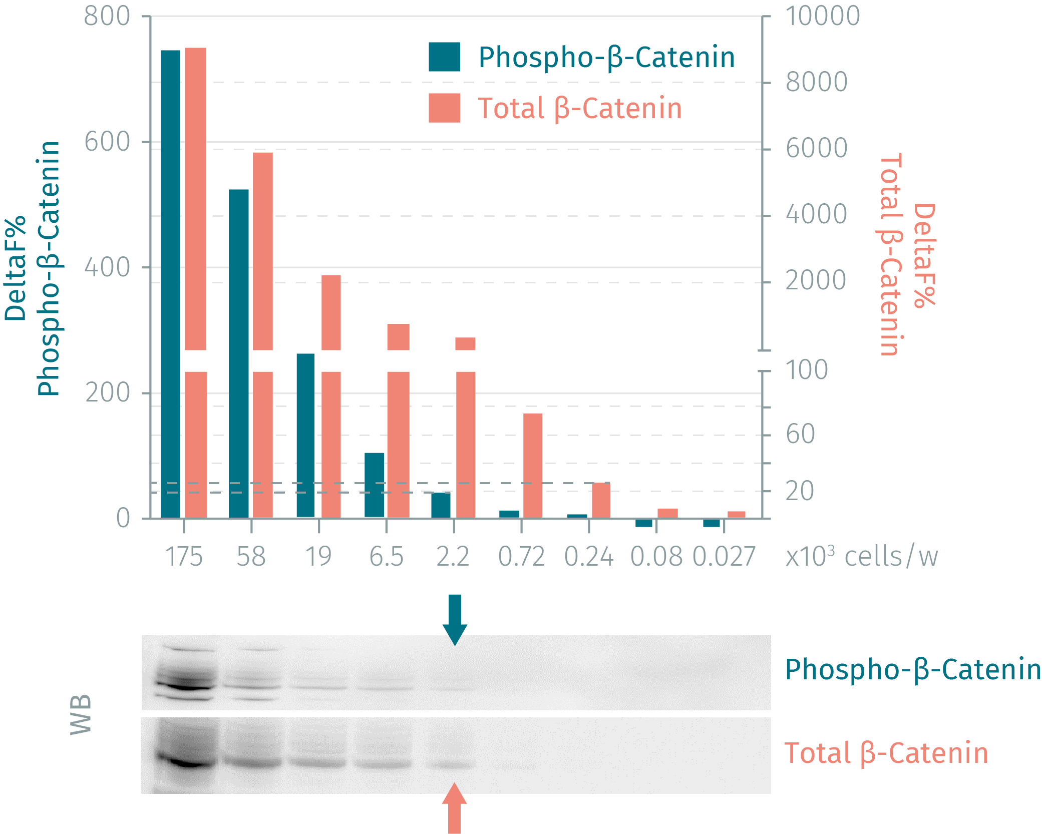
HTRF assay compared to Western blot using phospho-ß-Catenin and total ß-Catenin cellular assays
Human HEK293 cells were grown in a T175 flask at 37°C, 5% CO2 until 80% confluence and treated with MG132 at 5µM (3h). Cell culture medium was discarded, and 3 mL of supplemented lysis buffer were added for a 30 minutes incubation at room temperature. Soluble supernatants were collected after a 10 minute centrifugation. Serial dilutions of the cell lysate were performed in the supplemented lysis buffer and 16 µL of each dilution were dispensed and analyzed side-by-side by Western Blot and by HTRF. The HTRF phospho-ß-Catenin (T41/S33/S37) assay is as sensitive as WB, whereas HTRF total ß-Catenin is 9-fold more sensitive than the Western Blot.
BIO dose-response, a GSK3 inhibitor, on HEK293 cells using phospho-beta-Catenin and total beta-Catenin cellular assays
200,000 human HEK293 cells were plated in 96 well plates and incubated for 24h at 37 °C - 5% CO2. The cells were then treated with MG-132 and increasing concentrations of BIO for 3h. For cells lysis, 50µl of lysis buffer were added for 30min at RT under gentle shaking. 16 µL of lysate were transferred into a 384-well sv white microplate and 4 µL of the HTRF phospho-beta-Catenin and total beta-Catenin detection reagents were added. The HTRF signal was recorded after an overnight incubation.

Compatibility of the HTRF phospho-ß-Catenin cellular assay with different cell lines
Cells were plated at different cell densities in 96 well plates and incubated for 24h at 37 °C - 5% CO2. Medium was removed and cells were lysed with 50µl of lysis buffer for 30min at RT under gentle shaking. 16 µL of lysate were transferred into a 384-well sv white microplate and 4 µL of the HTRF phospho-ß-Catenin detection reagents were added. The HTRF signal was recorded after an overnight incubation. HTRF quantification of the ß-Catenin expression level is in good agreement with the literature (sadot et al, Journal of cell sciences 2002). The ß-catenin content is almost 2.5 higher in the SW-480 cells compared to HCT-116 cells and 15 higher in the SW-480 cells compared to HEK-293 cells.

Inhibition of proteasome activity by MG-132, measured by the HTRF phospho-ß-Catenin and total beta-Catenin cellular assays
200,000 human HEK293 cells were plated in 96 well plates and incubated for 24h at 37 °C - 5% CO2. The cells were then treated with increasing concentrations of MG-132 for different incubation time. For cells lysis, 50µl of lysis buffer were added for 30min at RT under gentle shaking. 16 µL of lysate were transferred into a 384-well sv white microplate and 4 µL of the HTRF phospho-beta-Catenin and total beta-Catenin detection reagents were added. The HTRF signal was recorded after an overnight incubation.


Simplified pathway
The role of ß-Catenin in the canonical Wnt pathway
In the absence of extracellular WNT ligands, cytosolic ß-Catenin is phosphorylated on Thr41/Ser37/Ser33, thereby targeting the protein for ubiquitination, degradation and inactivation. Binding of extracellular WNT ligands Frizzled receptor and/or the LDL receptor related proteins LRP 5 and 6, transduces their signal intracellularly via activation of disheveled DVL proteins. This results in the inactivation of the destruction complex, allowing cytosolic, non-phosphorylated ß-Catenin to accumulate and thereafter to translocate to the nucleus.

Resources
Guide
HTRF solutions, guide to major applications
This guide provides you an overview of HTRF applications in several therapeutic areas.
Guide
Protein degradation awareness in a single guide
An in-depth review of molecular and cellular pathways
The maintenance of proteostasis, the biological mechanisms that control the...

SDS, COAs, Manuals and more
Are you looking for technical documents for this product. We have housed them in a dedicated section., click on the links below to explore.


How can we help you?
We are here to answer your questions.

