Overview
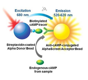
The AlphaScreen™ cAMP assay have been designed to directly measure levels of cAMP produced upon modulation of adenylate cyclase activity by GPCRs. The assay is based on the competition between endogenous cAMP and exogenously-added biotinylated cAMP. This is a displacement assay. The streptavidin-coated Donor bead will interact with the biotinylated cAMP tracer, and the Acceptor bead is coated with an anti-cAMP antibody to recognize both cellular and tracer cAMP. In the absence of cellular cAMP, the biotinylated cAMP antibody brings the Donor and Acceptor beads together, and excitation of the Donor bead results in emission of light from the Acceptor bead. In the presence of cellular cAMP (or cAMP from your sample), the Acceptor bead can bind to either the biotinylated cAMP probe or cAMP from your sample. The more cAMP in your sample, the more Acceptor bead that won't be available to bind the biotinylated cAMP probe, resulting in a decrease in signal. This assay can be used for screening or for quantitating the amount of cAMP in a sample by comparing it to a standard curve.

Figure 1. Principle of the AlphaScreen cAMP competition assay using a biotinylated cAMP tracer, a streptavidin-coated Donor bead, and an anti-cAMP antibody-coated Acceptor bead. Increasing amounts of intracellular cAMP will decrease the signal.
What do I need to run this assay?
Required reagents available from Revvity:
- AlphaScreen cAMP kit (catalog number 6760635D/M/R)
- Cells or cell membranes (or you can use your own cells/membranes)
- Microplates – We recommend our 96-well 1/2 AreaPlates or our 384-well white OptiPlates™.
- TopSeal™-A adhesive plate seal
Required reagents available from various suppliers (see where Revvityr R&D gets these reagents):
- Agonists/antagonists
- Forskolin (if working with Gi-coupled receptors)
- Buffer components for stimulation buffer (HEPES, Tween-20, BSA, PBS, HBSS)
- IBMX (phosphodiesterase inhibitor, to prevent degradation of cellular cAMP)
- Versene™ (to detach adherent cells gently)
- Ethanol
- DMSO for compound dilution
Instrumentation/equipment:
- A plate reader (capable of reading Alpha assays)
Alpha products and catalog numbers
View Alpha Products.
Protocol-in-brief
Please refer to the manuals for each kit, for detailed protocol information. As with all assay development, you may need to optimize the protocol for your application.
- Add 5 µL of cells + anti-cAMP Acceptor beads mix.
- Add 2.5 µL of antagonist.
- Add 2.5 µL of forskolin (if applicable)/agonist dilutions.
- Incubate in the dark for 30 minutes.
- Add 15 µL of biotinylated cAMP/streptavidin Donor beads detection mix.
- Incubate in the dark at room temperature for 4 hours.
- Read plate.
Assay optimization
- Forskolin dose response (if applicable)/cell titration
- Receptor stimulation time
- Agonist dose response
- Antagonist dose response
- Time course of cAMP detection
- Z' determination
Tips and FAQs
Q. What is the minimum receptor expression level required?
A. There is no minimum receptor expression level per se. Low levels of endogenous receptors, such as calcitonin receptors in CHO cells (15 fmol/mg), were shown to induce the release of high amounts of cAMP. Tight coupling between receptors, G-proteins (Gi or Gs), and adenylate cyclase is the most critical determinant to yield conditions for robust cAMP assays.
Q. Should I use transient or stable transfected cell lines?
A. Both transient and stable cell lines have been shown to produce good responses upon adenylate cyclase activation.
Q. How many cells should I use in my assay?
A. Cell number will influence both basal and stimulated cAMP levels. Performing a cell titration will allow you to optimize the signal window by maximizing the difference between the basal and stimulated counts.
Q. Is it necessary to perform a time course for cell stimulation?
A. The stimulation time is critical for reaching optimal detection of cAMP. When determining the optimal cell number for the assay, we recommend stimulating the cells for 30 minutes as a first trial. Once the optimal cell number has been determined, you should perform a time course experiment starting at 15 minutes up to 120 minutes, using 15-minute intervals. The time of stimulation may vary depending on the cell line, receptor, and agonist being studied.
Q. How should cells be handled?
A. Cells should be approximately 70-90% confluent and prepared just prior to the assay. Harvest at least 250,000 cells by detaching with PBS + 5 mM EDTA (Versene™ solution; Gibco catalog number 15040-066) for 5 min at 37°C. Centrifuge the cells for 5 min at 275 x g. Remove the supernatant and resuspend the cells in 1.5 mL of 1X PBS. Obtain an accurate cell count and adjust to 10^6 cells/mL with stimulation buffer. Cells should be >95% viable as determined by trypan blue staining.
Q. Can attached cells be used?
A. Attached cells can be used as well as detached cells. However, we recommend replacing the cell culture medium by the recommended stimulation buffer for at least 15 minutes prior to the assay. The high-level protocol would be:
Grow cells in a plate.
Remove the medium. Replace with a stimulation buffer containing agonists and/or compounds and IBMX.
Remove the medium. Lyse cells in the lysis buffer.
Transfer a fraction of the lysis buffer containing the lysed cells to a 96- or 384-well plate. Initially perform a titration to determine how much lysate to use.
Add other components in the lysis/detection buffer.
Incubate and read.
Q. Can membrane preparations be used instead of cells?
A. Membranes expressing Gs-coupled receptors were shown to produce excellent results when the stimulation buffer is supplemented with the appropriate ions (e.g., 10 mM MgCl2, 1 nM GTP, 10 µM GDP, and 100 µM ADP). 1-10 µg membranes are typically used in such assays. We recommend titering the membranes and all supplemented ions to optimize the performance of the assay.
Q. Does IBMX interfere with the assay?
A. IBMX is a well-known phosphodiesterase inhibitor. The inhibitory effect is due to its structural analogy to cAMP. Because of its resemblance to cAMP, IBMX competes with biotinylated cAMP binding to anti-cAMP antibodies on the Acceptor bead. High concentrations of IBMX decrease the Alpha signal without affecting assay sensitivity. We recommend using IBMX at a final concentration of 0.2-0.25 mM. At this concentration range, the maximum signal is reduced by approximately 30%.
For research use only. Not for use in diagnostic procedures.
The information provided above is solely for informational and research purposes only. Revvity assumes no liability or responsibility for any injuries, losses, or damages resulting from the use or misuse of the provided information, and Revvity assumes no liability for any outcomes resulting from the use or misuse of any recommendations. The information is provided on an "as is" basis without warranties of any kind. Users are responsible for determining the suitability of any recommendations for the user’s particular research. Any recommendations provided by Revvity should not be considered a substitute for a user’s own professional judgment.


