


Overview
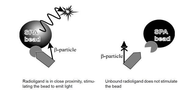
SPA is a versatile technology for the rapid and sensitive assay of a wide range of biological processes including the study of cellular receptor targets, enzyme function and molecular interactions between proteins and nucleic acids. The assay principle is depicted in its name: SPA beads or plates convert the energy from radioactive decay of β-particles when they come in close proximity to scintillant material embedded within the SPA bead, or base of a CytoStar-T™ plate. Blue light emission from the SPA scintillation beads or CytoStar-T plate is then detected in a PMT-based scintillation counter (e.g., MicroBeta2™). Red light emission from the SPA imaging beads is likewise detected in a CCD camera-based detector. By virtue of the energetics of the β- particles, radioactive decay that occurs in solution at a distance greater than the decay path length of the β- particle in aqueous solution will not stimulate the scintillant to emit light (Figure 1). This discrimination makes SPA a homogeneous assay format. Free ligands do not need to be physically separated from bound ligand for specific signal generation above background (good signal to noise ratio).
SPA is scalable from single tubes to 1536-well plates and is amenable to automation. A key benefit of this platform technology is that the biological molecules, ligands, or substrates being studied can be specifically labeled with either 3H, 125I, and 14C without perturbing the biology of the reaction being studied. SPA is well-suited for radiometric high-throughput screening. Applications include receptor-ligand binding assays, enzyme assays, radioimmunoassays, cell uptake assays, and molecular interaction assays.
Figure 1. Principle of the Scintillation Proximity Assay technology.
SPA beads and CytoStar-T plates: understanding the difference
Scintillation Proximity Assays can be performed in different formats: bead-based and plate-based. The choice of which format to use is best made on an application basis.
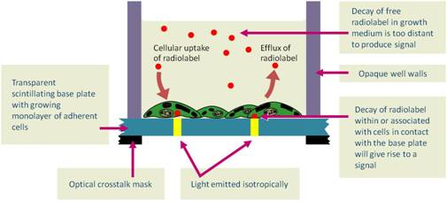
- CytoStar-T plates are recommended for cell-based radiometric detection assays. CytoStar-T plates are sterile, tissue-culture treated microplates that are specifically designed for non-invasive, live cell analysis in real-time following the principles of SPA.
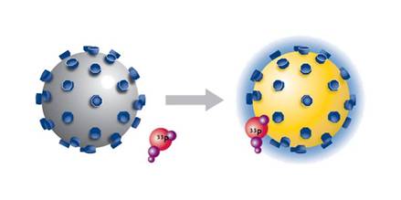
- For biochemical assays, including receptor-ligand binding, enzymatic, molecular interaction and homogeneous RIA, the bead-based format (SPA beads) should be used. SPA bead-based analyses can be done in single tubes, 96-, 384- and 1536-well microplate formats. The benefit of the bead-based format is that it provides the highest surface area for the biological reaction to occur and provides the flexibility to study the interaction either ON-bead or OFF-bead.
Scintillation proximity technologies
SPA beads
View more information on SPA bead technology and applications. Scintillation proximity assay (SPA) is a homogeneous and versatile technology for the rapid and sensitive assay of a wide range of biological processes, including applications using enzyme and receptor targets, radioimmunoassays, and molecular interactions. When 3H, 14C, and 125I radioisotopes decay, they release β-particles (or Auger electrons, in the case of 125I). The distance these particles travel through an aqueous solution is dependent on the energy of the particle. If a radioactive molecule is held in close enough proximity to a SPA Scintillation Bead or a SPA Imaging Bead, the decay particles stimulate the scintillant within the bead to emit light, which is then detected in a PMT-based scintillation counter or on a CCD-based imager respectively. However, if the radioactive molecule does not associate with the SPA bead, the decay particles will not have sufficient energy to reach the bead and no light will be emitted. This discrimination of binding by proximity means that no physical separation of bound and free radiochemical is required.
SPA beads
CytoStar-T plates
View more information on CytoStar-T technology and applications. This plate-based version of scintillation proximity assays utilizes sterile, tissue-culture treated microplates. CytoStar-T scintillating microplates are sterile, tissue culture treated microplates designed not only for the growth of adherent but also suspension cell cultures. The integral, planar, transparent base of each well is composed of a proprietary homogeneous mixture of scintillant and polystyrene. The transparent nature of the base permits the observation of growth of cells plated in the well. Radioisotopes having suitable decay characteristics (3H, 14C, 35S, 125I) brought into proximity with the scintillant contained within the base (via radiochemical uptake or radiochemical interaction with the cells) will have that radioactive decay converted to a light signal. The amount of light generated is proportional to the amount of radioisotope within, or associated with, the cells.
CytoStar-T technology
Custom services
Revvity offers several custom radiochemical services. If you are interested in custom services, please contact our custom teams.
Radiosynthesis and Labeling Custom Services
For research use only. Not for use in diagnostic procedures.
The information provided above is solely for informational and research purposes only. Revvity assumes no liability or responsibility for any injuries, losses, or damages resulting from the use or misuse of the provided information, and Revvity assumes no liability for any outcomes resulting from the use or misuse of any recommendations. The information is provided on an "as is" basis without warranties of any kind. Users are responsible for determining the suitability of any recommendations for the user’s particular research. Any recommendations provided by Revvity should not be considered a substitute for a user’s own professional judgment.