


Overview
Neutrophil elastase is a key protease involved in acute lung injury, acute respiratory distress syndrome, as well as many other inflammatory processes such as emphysema, cystic fibrosis, COPD, wound healing, rheumatoid arthritis, and ischemia-reperfusion. IVISense™ Neutrophil Elastase 680 FAST is a fluorescent probe that is selectively cleaved by elastase produced by activated neutrophils during acute inflammation. It is optically silent upon injection and produces fluorescent signal only after cleavage by elastase.
IVISense Neutrophil Elastase 680 FAST probe is a member of a family of activatable fluorescent imaging probes comprising a novel architecture, termed F.A.S.T. (Fluorescent Activatable Sensor Technology) that confers an improved pharmacokinetic profile with a broader range of early imaging time points. This architecture also offers higher target specific signal with reduced background. It can be used for imaging both in vitro and in vivo.
IVISense Neutrophil Elastase 680 FAST probe enables imaging of neutrophil elastase activity in applications including:
- Acute lung injury models
- Acute respiratory distress syndrome
- Emphysema
- Cystic Fibrosis
- COPD
- Wound Healing
- Rheumatoid Arthritis
- Ischemia-reperfusion
Products and catalog numbers
| Product | Catalog Number | Ex/Em wavelength (nm) | Molecular weight (g/mol) | Validated Experiments | Applications |
|---|---|---|---|---|---|
| IVISense Neutrophil Elastase 680 FAST | NEV11169 | 675/693 | 43,000 | In vivo/Ex vivo | Acute inflammation Pulmonary Diseases |
Using IVISense Neutrophil Elastase 680 FAST probe for in vivo/ex vivo studies
The generally recommended procedure for in vivo imaging with IVISense Neutrophil Elastase 680 FAST probe is administration via intravenous injection and imaging 4-8 hours post injection. Earlier and later time points may be appropriate for some disease models, and the optimal imaging time point for any application should be determined empirically.
View instructions on setting up an in vivo mouse experiment with IVISense Neutrophil Elastase 680 FAST fluorescent probe
| Product | Route of Injection | Mouse Dose (25 g) | Rat Dose (250 g) | Blood t 1/2 | Tissue t 1/2 | Optimal imaging time | Optimal Re-injection Time (complete clearance) | Route of Metabolism/ background tissue | FMT& IVIS settings |
|---|---|---|---|---|---|---|---|---|---|
| IVISense Neutrophil Elastase 680 FAST | IV | 4 nmol | 12-40 nmol | 3 h | 12 h | 3-6 h | 2 d | Bladder, liver, intestines | FMT 680/700 IVIS 675/720 |
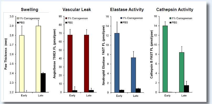
Figure 1: Paw inflammation was induced with carageenan (CG) injection. CG-injected mice were measured for changes in paw thickness, vascular leak (IVISense Vascular 750 Fluorescent Probe), neutrophil elastase activity (IVISense Neutrophil Elastase 680 FAST Fluorescent Probe), and cathepsin activity (IVISense Cat B 680 FAST Fluorescent Probe) by injecting these probes at two time points, 2h (early) and 24h (late), post CG injection. Tomographic (3D) paw imaging was performed on the FMT2500 5h after probe injection. All CG-induced responses were statistically significant at both time points (p < 0.01), revealing significant recruitment of inflammatory cells.
Frozen Tissue Protocol
We have validated IVISense Neutrophil Elastase 680 FAST for use with frozen tissue samples. Here is a brief protocol with a recommended concentration of probe to use:
- Freeze tumor (without probe) and section 5-10 µm by cryostat. For lung samples, mice are challenged intranasally with 100 µg of LPS followed by intranasal instillation of 200 nM fMLP in 40 µL PBS 18 h later. Harvest lungs 5 h later. For specificity, co-incubate probe with Silvelestat inhibitor.
- Incubate with 1 µM IVISense Neutrophil Elastase 680 FAST at 37ºC for 10-30 min for tumors; 5 h for lungs.
- Wash 1x with PBS.
- Mount with anti-fade reagent.
- Fluorescence microscopy filter: Cy5.5
Application notes and posters
- Poster: A novel NIR dye for in vivo temporal tracking of labeled macrophages to sites of acute inflammation
- Poster: Near-Infrared Quantitative Fluorescence Imaging of Renin Activity in Kidney Tissue
For research use only. Not for use in diagnostic procedures. The information provided above is solely for informational and research purposes only. The information does not constitute medical advice and must not be used or interpreted as such. Consult a qualified veterinarian or researcher for specific guidance or use information. Revvity assumes no liability or responsibility for any injuries, losses, or damages resulting from the use or misuse of the provided information, and Revvity assumes no liability for any outcomes resulting from the use or misuse of any recommendations. The information is provided on an "as is" basis without warranties of any kind. Users are responsible for determining the suitability of any recommendations for the user’s particular research. Any recommendations provided by Revvity should not be considered a substitute for a user’s own professional judgment. Users are solely responsible for complying with all relevant laws, regulations, and institutional animal care and use committee (IACUC) guidelines in their use of the information provided.