
PhenoVue Multi Organelle Staining Kit 1x384
 View All
PhenoVue Multi Organelle Staining Kit 1x384
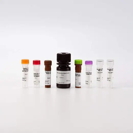
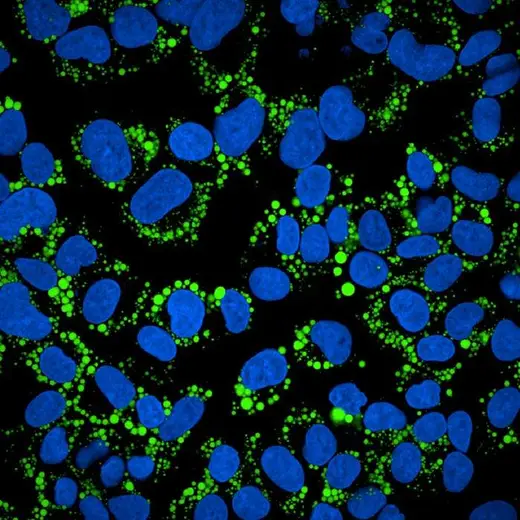
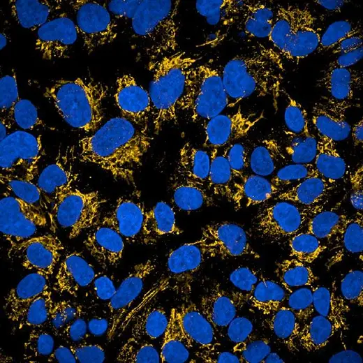
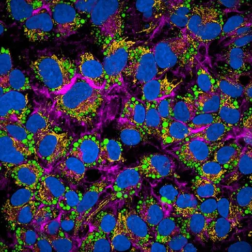
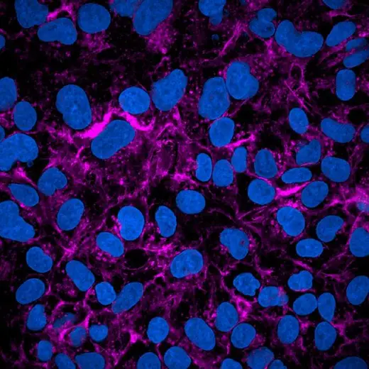
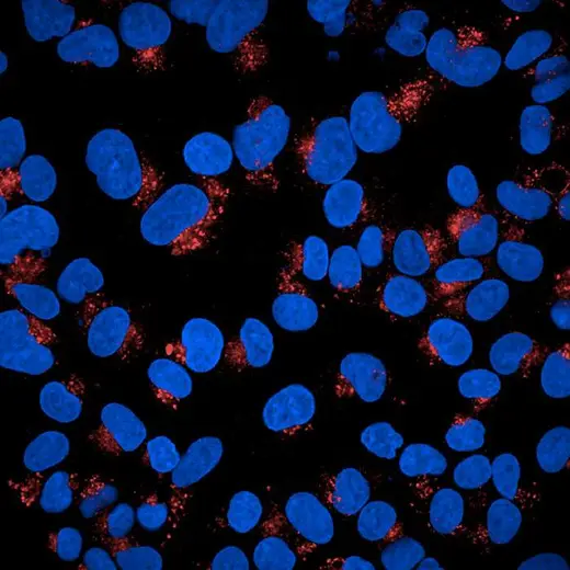


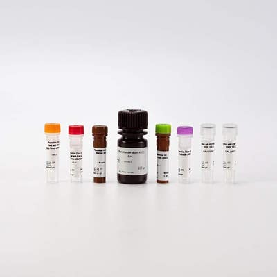
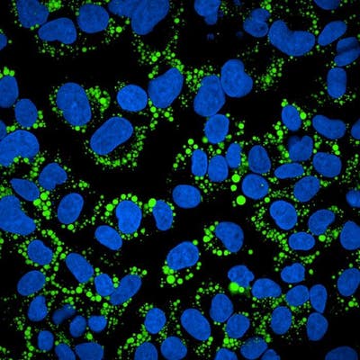
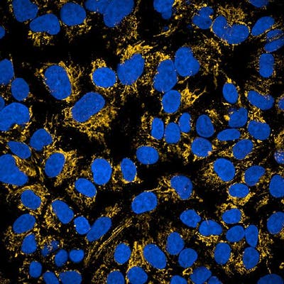
PhenoVue™ Multi Organelle Staining Kit comprises five validated, optimized and ready-to-use fluorescent probes to visualize mitochondria, lysosomes, lipid droplets, actin cytoskeleton, and nuclei in a 5-plex experiment.
The kit is validated for use on high-content screening systems such as the Opera Phenix® Plus high content screening system, and is ideal for the study of metabolic disorders, infectious diseases, and liver toxicity.
View our extensive validation data in the Product Information Sheet within the Resources tab below.
For research use only. Not for use in diagnostic procedures.


PhenoVue Multi Organelle Staining Kit 1x384


PhenoVue Multi Organelle Staining Kit 1x384


Product information
Overview
PhenoVue Multi Organelle Staining Kit includes five PhenoVue probes to visualize nuclei, mitochondria, lysosomes, actin, and lipid droplets. These organelles play crucial roles in various cellular processes and interact with each other in intricate ways to maintain cell homeostasis and carry out specific cellular processes.
The combination of specific, bright and photostable PhenoVue dyes enables the ability to perform a 5-plex experiment with maximum spectral separation and a significantly reduced spectral overlap.
Each PhenoVue probe has been extensively validated and carefully optimized for excellent batch-to-batch reproducibility.
The PhenoVue Multi Organelle Staining Kit offers a straightforward and user-friendly protocol to facilitate the experimental procedure. Avoiding the use of living cells, it provides a convenient alternative method for phenotypic screening approaches such as the well-established cell painting protocol.
Kit Components / Quantity:
- PhenoVue Hoechst 33342 Nuclear Stain / 1 vial of 70 µL (50000x)
- PhenoVue 493 Lipid Stain / 1 vial of 30 µL (4000x)
- PhenoVue Fluor 400LS - Phalloidin / 1 vial of 30 µL (400X)
- PhenoVue anti-HSP60 antibody 100X / 1 vial of 100 µL (100x)
- PhenoVue anti-LAMP1 antibody 100X / 1 vial of 100 µL (100x)
- PhenoVue Fluor 555 - Goat Anti-Mouse Antibody Highly Cross-Adsorbed / 1 vial of 100 µL (100x)
- PhenoVue Fluor 647 - Goat Anti-Rat Antibody Highly Cross-Adsorbed / 1 vial of 50 µL (200x)
- PhenoVue Dye Diluent A (5X) / 1 vial - 8 mL (5X)
Specifications
| Application |
High Content Imaging
Microscopy
|
|---|---|
| Assay Points |
1 x 384-well microplate
|
| Brand |
PhenoVue™
|
| Detection Modality |
Fluorescence
|
| Organelle and Cell Compartment |
Actin
Lipid droplets
Lysosomes
Mitochondria
Nuclei
|
| Quantity |
1 kit
|
| Sample Type |
Fixed samples only
|
| Shipping Conditions |
Shipped in Dry Ice
|
| Storage Conditions |
-16 °C or below, protected from light
|
| Type |
Kit
|
Image gallery


PhenoVue Multi Organelle Staining Kit 1x384


PhenoVue Multi Organelle Staining Kit 1x384


Spectra Viewer
Resources
Are you looking for resources, click on the resource type to explore further.
This flyer describes Revvity's PhenoVue cellular imaging reagents.


How can we help you?
We are here to answer your questions.


