PhenoVue Fluorescent Probes for Organelles & Subcellular Compartments
Optimized for high-content screening and analysis
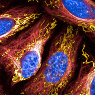
Cellular imaging techniques, such as high-content analysis, rely on the ability to detect and distinguish between specific cellular compartments and organelles. High-quality data depends on high-quality images, which all depends on bright fluorescent dyes. Building on our extensive expertise in imaging instrumentation, fluorescent dye chemistry, and assay development, our PhenoVue organelle-specific stains detect various organelles and cellular compartments and are optimized for high-content analysis and screening. These fluorescent stains can be used for cell membrane, endoplasmic reticulum, golgi apparatus, lipids, lysosomes, nucleoli, nuclei, mitochondria, actin cytoskeleton, and tubulin. For convenience, our multi organelle staining kit contains five ready to use bright fluorescent probes to visualize five key organelles. Applicable to fixed cell models, this kit provides an alternative to the established cell painting assay.
Cell compartment and organelle stains:
- Cell membrane/endoplasmic reticulum/golgi apparatus: PhenoVue Fluor – WGA and PhenoVue Fluor - Concanavalin A
- Actin cytoskeleton: PhenoVue Fluor – Phalloidin and PhenoVue Fluor Live Cell Actin Stain
- Tubulin network: PhenoVue Fluor Live Cell Tubulin Stains
- Lysosomes: PhenoVue Lysosomal Stains
- Lipid droplets: PhenoVue Nile Red and PhenoVue 493 Lipid Stain
- Nucleoli: PhenoVue 512 Nucleic Acid Stain
- Nuclei: PhenoVue Hoechst 33342 Nuclear Stain, PhenoVue DAPI Nuclear Stain, PhenoVue DRAQ7™ Dead Cell Nuclear Stain, and PhenoVue DRAQ5™ Total Cell Nuclear Stain
- Mitochondria: PhenoVue Mitochondrial Stains
Features include:
- Multi organelle staining kit and neuronal differentiation staining kit to streamline your workflow
- Range of stains and fluors for commonly studied organelles and compartments
- Variety of fluorescent colors to enable multiplexing while avoiding spectral overlap
- Validation in high-content screening applications
- Bright fluorophores to enable high-quality images
For research use only. Not for use in diagnostic procedures.


