


US
Revvity Sites Globally
Select your location.
*e-commerce not available for this region.

HTRF Human Phospho-ZAP70 (Tyr319) Detection Kit, 500 Assay Points


HTRF Human Phospho-ZAP70 (Tyr319) Detection Kit, 500 Assay Points


Shipping box for Revvity reagent kits


The phospho-ZAP-70 (Tyr319) assay enables the cell-based quantitative detection of ZAP-70 phosphorylated on Ser Tyr319 as a readout of T-cell activation.
| Feature | Specification |
|---|---|
| Application | Cell Signaling |
| Sample Volume | 16 µL |
The phospho-ZAP-70 (Tyr319) assay enables the cell-based quantitative detection of ZAP-70 phosphorylated on Ser Tyr319 as a readout of T-cell activation.
Product Variants
Unit Size: 500 Assay Points
Part #:
64ZAPPEG
List Price
USD 2,271.53
Unit Size: 10,000 Assay Points
Part #:
64ZAPPEH
List Price
USD 13,214.42
For research use only. Not for use in diagnostic procedures. All products to be used in accordance with applicable laws and regulations including without limitation, consumption and disposal requirements under European REACH regulations (EC 1907/2006).
HTRF Human Phospho-ZAP70 (Tyr319) Detection Kit, 500 Assay Points
Shipping box for Revvity reagent kits
HTRF Human Phospho-ZAP70 (Tyr319) Detection Kit, 500 Assay Points
Product information
Overview
The Phospho-ZAP-70 (Tyr319) cellular assay is ideal for monitoring T-cell activation, which results in the phosphorylation of ZAP-70 on Tyrosine 319. ZAP-70 is a cytoplasmic protein tyrosine kinase expressed in T and NK cells, and plays a critical role in the events involved in initiating T-cell responses by the antigen receptor.
Specifications
| Application |
Cell Signaling
|
|---|---|
| Brand |
HTRF
|
| Detection Modality |
HTRF
|
| Lysis Buffer Compatibility |
Lysis Buffer 3
Lysis Buffer 4
Lysis Buffer 5
|
| Molecular Modification |
Phosphorylation
|
| Product Group |
Kit
|
| Sample Volume |
16 µL
|
| Shipping Conditions |
Shipped in Dry Ice
|
| Target Class |
Phosphoproteins
|
| Target Species |
Human
|
| Technology |
TR-FRET
|
| Therapeutic Area |
Oncology & Inflammation
|
| Unit Size |
500 Assay Points
|
Video gallery
HTRF Human Phospho-ZAP70 (Tyr319) Detection Kit, 500 Assay Points
HTRF Human Phospho-ZAP70 (Tyr319) Detection Kit, 500 Assay Points
How it works
Phospho-ZAP-70 (Tyr139) assay principle
The Phospho-ZAP-70 (Tyr139) assay measures ZAP-70 when phosphorylated at Tyr139. Contrary to Western Blot, the assay is entirely plate-based and does not require gels, electrophoresis or transfer. The Phospho-ZAP-70 (Tyr139) assay uses 2 labeled antibodies: one with a donor fluorophore, the other one with an acceptor. The first antibody is selected for its specific binding to the phosphorylated motif on the protein, the second for its ability to recognize the protein independent of its phosphorylation state. Protein phosphorylation enables an immune-complex formation involving both labeled antibodies and which brings the donor fluorophore into close proximity to the acceptor, thereby generating a FRET signal. Its intensity is directly proportional to the concentration of phosphorylated protein present in the sample, and provides a means of assessing the proteins phosphorylation state under a no-wash assay format.
Phospho-ZAP-70 (Tyr139) 2-plate assay protocol
The 2 plate protocol involves culturing cells in a 96-well plate before lysis then transferring lysates to a 384-well low volume detection plate before adding phospho-ZAP-70 (Tyr139) HTRF detection reagents. This protocol enables the cells' viability and confluence to be monitored.

Phospho-ZAP-70 (Tyr139) 1-plate assay protocol
Detection of Phosphorylated ZAP-70 (Tyr139) with HTRF reagents can be performed in a single plate used for culturing, stimulation and lysis. No washing steps are required. This HTS designed protocol enables miniaturization while maintaining robust HTRF quality.

Assay validation
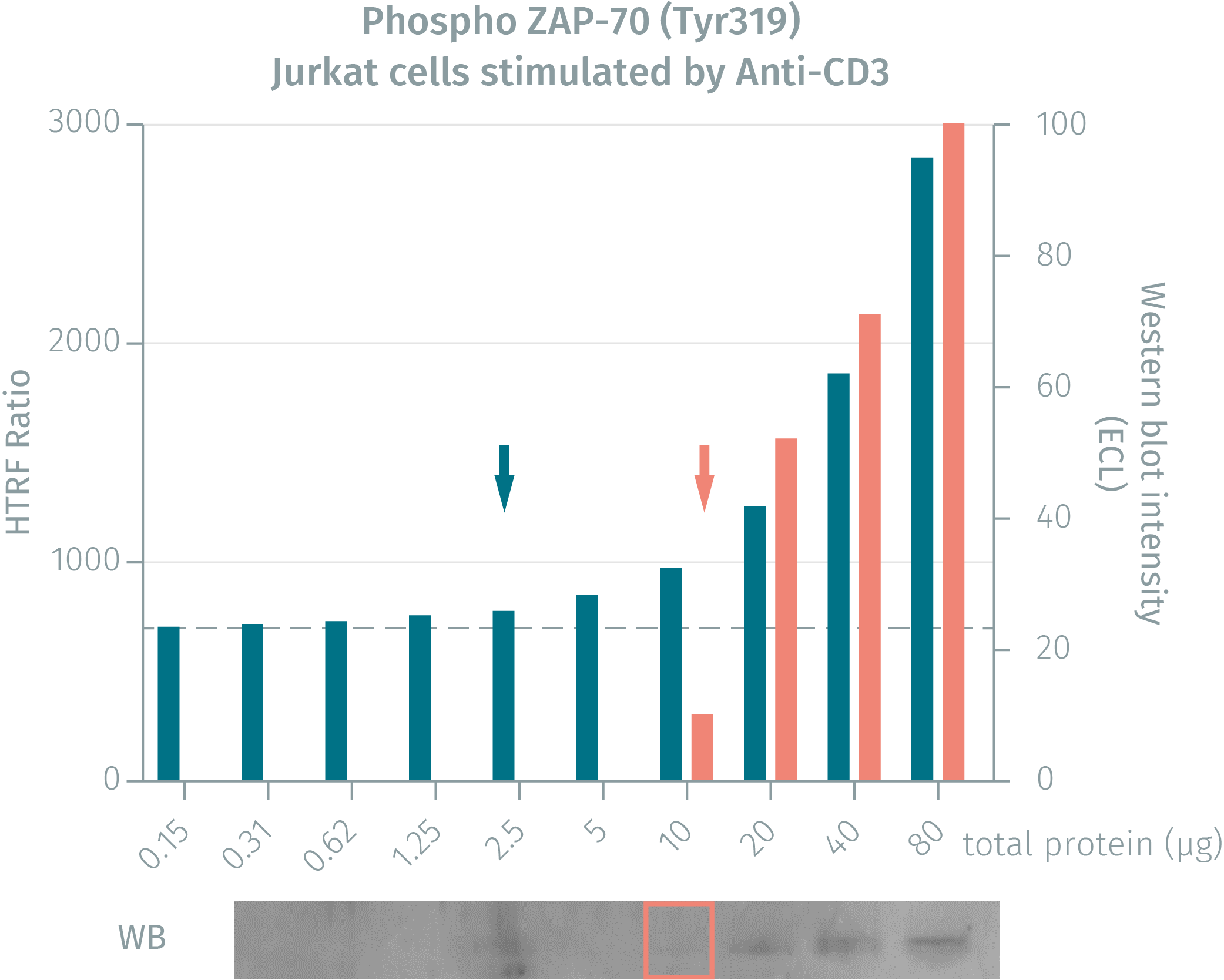
HTRF assay compared to Western Blot using phospho-ZAP-70 (Tyr319) cellular assays on human Jurkat cells
Jurkat cells were grown in a T175cm2 flask for 2 days in RPMI culture medium supplemented by 10% FBS, at 37°C in 5% CO2 atmosphere. 4.5 ml of cell suspension (13 x 106 cells / mL) were stimulated with anti-CD3 antibody (20 µg / mL final concentration during 2.5min). After stimulation, cells were lysed at RT for 30 min by adding 2.25 mL of 4X supplemented lysis buffer. Neat lysate was then serially diluted in the same supplemented lysis buffer. 16 µL of each dilution were analyzed in parallel by HTRF or Western Blot
Cell density optimisation for Phospho ZAP-70 (Tyr319)
This assay was performed using the two-plate assay protocol. Jurkat cells were plated in a 96-well culture plate at 200K cells or 400K cells and 25 µL / well, in complete RPMI culture medium with 10% FBS. Cell stimulation was done by anti-CD3 Ab at the final concentration of 20 µg / mL for 2.5 min. Cells lysis was performed using 10 µL of 4X supplemented lysis buffer. 16 µL of each cell lysate were transferred to the detection plate for analysis using HTRF Phospho ZAP-70 (Tyr319) assays.

Kinetics of cell stimulation - Phospho ZAP-70 (Tyr319) assay
This assay was performed using the two-plate assay protocol. Jurkat cells were plated in a 96-well culture plate at 400 K cells, using 25 µL / well in complete RPMI culture medium with 10% FBS. Cell stimulation was performed using the anti-CD3 Ab at the final concentration of 20 µg / mL for different stimulation times (min). After stimulation, cells were lysed with 10 µL of 4X supplemented lysis buffer. 16 µl of each cell lysate type were analyzed with the HTRF phospho ZAP-70 (Tyr319) assay.

Monitoring of the immune checkpoint inhibitor PD-L1 activity
As Phospho ZAP-70 (Tyr319) is a readout of T cell activation, it was used to monitor the inhibitory effect of PD-L1 recombinant protein on T cell activation. This assay was performed using the two-plate assay protocol. 400 000 Jurkat cells were plated in 25 µL of complete RPMI culture medium with 10% FBS in a 96-well culture plate. Cells were pre-incubated 24 hours with different concentrations of PD-L1 recombinant protein. Cells were then stimulated for 2.5 min with 20 µg / mL anti-CD3 Ab (final concentration) and lysed directly with10 µL of 4X supplemented lysis buffer, then incubated 30 min at RT. 16µL of each sample were analyzed using Phospho ZAP-70 (Tyr319) and Total ZAP-70 assays. Phospho ZAP-70 results were normalized with the Total ZAP-70 amount and plotted on a graph. A slight but significant inhibitory effect of PD-L1 recombinant protein was recorded with the Phospho ZAP-70 (Tyr319) assay at the final concentration of 40 µg / mL.

Simplified pathway
Function and regulation of ZAP-70
TCR engagement promotes the phosphorylation of immunoreceptor tyrosine-based activation motifs (ITAMs) on the cytosolic side of the TCR/CD3 complex by lymphocyte protein tyrosine kinase (Lck). Zap-70 is recruited to the TCR/CD3 complex where it becomes phosphorylated and activated, promoting recruitment and phosphorylation of downstream adaptor or scaffold proteins.

Resources
Are you looking for resources, click on the resource type to explore further.
Brochure
HTRF assays and reagents catalog
Discover the versatility and precision of Homogeneous Time-Resolved Fluorescence (HTRF) technology. Our HTRF portfolio offers a...
Guide
HTRF solutions, guide to major applications
This guide provides you an overview of HTRF applications in several therapeutic areas.


How can we help you?
We are here to answer your questions.

