
HTRF Human & Mouse TGF-β1 Detection Kit, 10,000 Assay Points


HTRF Human & Mouse TGF-β1 Detection Kit, 10,000 Assay Points


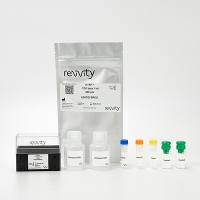
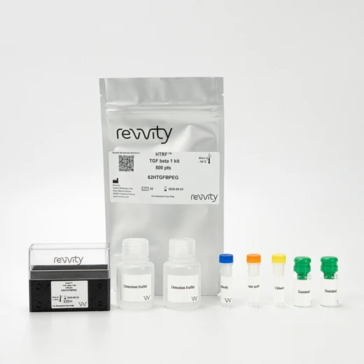
The HTRF human and mouse TGF beta 1 kit is designed for the quantification of human or mouse TGF beta 1 release in cell supernatant.
For research use only. Not for use in diagnostic procedures. All products to be used in accordance with applicable laws and regulations including without limitation, consumption and disposal requirements under European REACH regulations (EC 1907/2006).
| Feature | Specification |
|---|---|
| Application | Protein Quantification |
| Sample Volume | 16 µL |
The HTRF human and mouse TGF beta 1 kit is designed for the quantification of human or mouse TGF beta 1 release in cell supernatant.
For research use only. Not for use in diagnostic procedures. All products to be used in accordance with applicable laws and regulations including without limitation, consumption and disposal requirements under European REACH regulations (EC 1907/2006).


HTRF Human & Mouse TGF-β1 Detection Kit, 10,000 Assay Points


HTRF Human & Mouse TGF-β1 Detection Kit, 10,000 Assay Points


Product information
Overview
TGF beta 1 (Transforming Growth Factor Beta-1) is considered one of major cytokines involved in regulation of extracellular matrix (ECM) synsis and degradation, as well as a major profibrotic factor. TGF beta 1 pre-proprotein is processed to generate a latency-associated peptide (LAP) and a mature TGFß1 peptide, which remain associated through strong non-covalent interactions. An activation step (MMP2 cleavage or acid activation) is required to release active form of TGF beta 1 from TGFß1-LAP complex.
Assessment of serum samples often requires enhanced sensitivity. In some cases, AlphaLISA assays may have sufficient sensitivity to enable detection of low levels of analytes in serum or plasma. When assaying, always follow recommended protocol and avoid highly haemolyzed samples.
Specifications
| Application |
Protein Quantification
|
|---|---|
| Brand |
HTRF
|
| Detection Modality |
HTRF
|
| Product Group |
Kit
|
| Sample Volume |
16 µL
|
| Shipping Conditions |
Shipped in Dry Ice
|
| Target Class |
Cytokines
|
| Target Species |
Human
Mouse
|
| Technology |
TR-FRET
|
| Therapeutic Area |
Metabolism/Diabetes
NASH/Fibrosis
Oncology & Inflammation
|
| Unit Size |
10,000 Assay Points
|
Video gallery

HTRF Human & Mouse TGF-β1 Detection Kit, 10,000 Assay Points

HTRF Human & Mouse TGF-β1 Detection Kit, 10,000 Assay Points

How it works
Activation step
The samples and cell culture supernatants contain TGF beta 1 and a TGFß1-LAP complex, which require an acid activation step followed by a neutralization in order to be detected (sample activation can follow one of the protocols described on the right). The reagents needed for this activation step are provided in the kit for maximum convenience. Standards and activated samples are dispensed directly into the detection (white, low-volume) plate for the detection by HTRF® reagents.

Assay principle
Cell supernatant, sample, or standard is dispensed directly into the assay plate for the detection by HTRF® reagents (384-well low-volume white plate or Revvity low-volume 96-well plate in 20 µl). The antibodies labeled with the HTRF donor and acceptor are pre-mixed and added in a single dispensing step, to further streamline the assay procedure. The assay can be run up to a 1536 well-format by simply resizing each addition volume proportionally.

Assay details
Technical specifications of human and mouse TGF beta 1 kit
| Sample size | 16 µL |
|---|---|
| Final assay volume | 20 µL |
| Kit components | Lyophilized standard, frozen detection antibodies, buffers &protocol |
| LOD &LOQ (in Diluent) | 4 pg/mL &19 pg/mL |
| Range | 19 2,000 pg/mL |
| Time to result | ON at RT |
| Calibration | NIBSC (89/514) value (U/mL) = 0.017 x HTRF hTGFß value (pg/mL) |
| Species | Human, mouse, bovine (use of FCS is possible up to 5% max), others expected (based on sequences similarities). No detection of chicken TGFß1. |
| Specificity | There is no cross-reactivity with human TGFß2 and TGFß3. |
Analytical performance
Intra and inter assay
Intra assay (n=24)
| Sample | Mean [TGFβ1] (pg/mL) | CV |
|---|---|---|
| 1 | 28 | 7% |
| 2 | 167 | 8% |
| 3 | 704 | 4% |
| Mean CV | 6% |
Inter assay (n=4)
| Sample | [TGFβ1] (pg/mL) | Mean (delta ratio) | CV |
|---|---|---|---|
| 1 | 32 | 114 | 3% |
| 2 | 126 | 410 | 6% |
| 3 | 322 | 1441 | 2% |
| Mean CV | 4% |
Assay validation
Human TGFß1 secretion in PBMC cells stimulated with Ionomycin & PMA
Human PBMCs plated at 125 kcells/well (96 wells plate) in RPMI containing 4% FCS were stimulated for 18 h with Ionomycin (1 µg/mL) and PMA ranging from 0.5 to 50 ng/mL. After transfer into polypropylene microtubes, 50 µL of supernatants were treated with the acid activation reagent (5 µL, 10 min, room temperature) then with the neutralization reagent (5 µL). 16 µL were transferred into a white detection plate. Recommended controls include complemented media alone and unstimulated cells in complemented medium to determine TGF beta in stimulation media and baseline concentrations respectively.

Mouse TGFß1 secretion from immortalized kupffer cells
Immortalized mouse Kupffer cells (ImKC) were plated at 650 kcells/well (24 wells plate) in RPMI containing 4% FCS. After 24 h resting, the cells were stimulated for 16 h with increasing concentrations of LPS ranging from 0.05 to 5 µg/mL (final volume 500 µL/well). 100 µL of supernatants were acid activated (10 µL) for 10 min at room temperature then neutralized (10 µL). 16 µL of activated supernatants were then transferred into a white detection plate (384 low volume) to be analyzed by the Human TGF beta 1 Assay.

Resources
Are you looking for resources, click on the resource type to explore further.
Advance your autoimmune disease research and benefit from Revvity broad offering of reagent technologies


How can we help you?
We are here to answer your questions.


