


US
Search Results
Revvity Sites Globally
Select your location.
*e-commerce not available for this region.

HTRF Human and Mouse Phospho-SMAD3 (Ser423/425) Detection Kit, 10,000 Assay Points


Smad3 P-s423/425 Kit - 50,000 Tests
For research use only. Not for use in diagnostic procedures. All products to be used in accordance with applicable laws and regulations including without limitation, consumption & disposal requirements under European REACH regulations (EC 1907/2006).
Product Variants
Product information
Overview
This HTRF cell-based assay enables the rapid, quantitative detection of Phosphorylated SMAD3 phosphorylated at Serine 423/425 as a readout of TGF-ß signaling activity. TGF-ß receptors directly activate SMAD3 by phosphorylation at Ser423/425 causing it to translocate to the nucleus and regulate gene expression involved in apoptosis, migration and differentiation, as well as in immune/inflammatory responses and extracellular matrix remodeling.
Specifications
| Assay Points |
10000
|
|---|---|
| Assay Target Type |
Kit
|
| Assay Technology |
HTRF
|
| Brand |
HTRF
|
| Quantity |
1
|
| Therapeutic Area |
Cardiovascular
Metabolism/Diabetes
NASH/Fibrosis
Oncology & Inflammation
|
| Unit Size |
10,000 Assay Points
|
Video gallery
How it works
Phospho-SMAD3 (Ser423/425) assay principle
The Phospho-SMAD3 (Ser423/425) assay measures SMAD3 when phosphorylated at Ser423/425. Contrary to Western Blot, the assay is entirely plate-based and does not require gels, electrophoresis or transfer. The Phospho-SMAD3 (Ser423/425) assay uses 2 labeled antibodies: one with a donor fluorophore, the other one with an acceptor. The first antibody is selected for its specific binding to the phosphorylated motif on the protein, the second for its ability to recognize the protein independent of its phosphorylation state. Protein phosphorylation enables an immune-complex formation involving both labeled antibodies and which brings the donor fluorophore into close proximity to the acceptor, thereby generating a FRET signal. Its intensity is directly proportional to the concentration of phosphorylated protein present in the sample, and provides a means of assessing the proteins phosphorylation state under a no-wash assay format.
Phospho-SMAD3 (Ser423/425) 2-plate assay protocol
The 2 plate protocol involves culturing cells in a 96-well plate before lysis then transferring lysates to a 384-well low volume detection plate before adding Phospho-SMAD3 (Ser423/425) HTRF detection reagents. This protocol enables the cells' viability and confluence to be monitored.

Phospho-SMAD3 (Ser423/425) 1-plate assay protocol
Detection of Phosphorylated SMAD3 (Ser423/425) with HTRF reagents can be performed in a single plate used for culturing, stimulation and lysis. No washing steps are required. This HTS designed protocol enables miniaturization while maintaining robust HTRF quality.

Assay validation
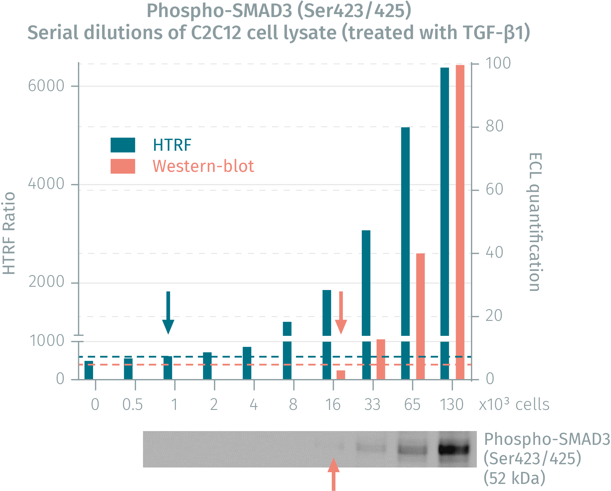
HTRF assay compared to Western Blot using phospho-SMAD3 assay
Cells were cultured to 80% confluency and treated with TGF-ß1 for 30 minutess. Following lysis, soluble supernatants were collected via centrifugation. Serial dilutions of ysates were performed and transferred into a low volume white microplate before finally adding HTRF phospho-SMAD3 detection reagents. Equal amounts of lysates were used for a side by side comparison between HTRF and Western Blot. The results reveal that the HTRF phospho-SMAD3 assay is 16-fold more sensitive than the Western Blot.
Validation on the mouse cell lines NIH/3T3 and C2C12
Mouse NIH/3T3 and C2C12 lines were plated (100,000 cells/well) and cultured 2h in complete culture medium, 37°C - 5% CO2. The day after, the cells were treated with increasing concentrations of TGF-ß1 for 30 minutes at 37°C - 5% CO2. After medium removal, the cells were lysed with 50 µL of supplemented lysis buffer #4 for 30 minutes at RT under gentle shaking, and 16 µL of lysate were transferred twice over into a low volume white microplate before adding 4 µL of the HTRF phospho-SMAD3 or total SMAD3 detection antibodies. The HTRF signal was recorded after an overnight incubation. In both lines, TGF-ß1 promotes the activation of SMAD3 by phosphorylation on Ser423/425, whereas the expression level of the protein remains fairly stable.


Overactivation of TGF-ß/SMAD signaling in hepatocytes and HSCs promotes liver fibrosis
Human LX-2* and HepG2 lines were plated (50,000 and 200,000 cells/well respectively) and cultured overnight in complete culture medium, 37°C - 5% CO2. Cells were then treated with increasing concentrations of TGF-ß1 for 30 minutes at 37°C - 5% CO2. After medium removal, cells were lysed with 50 µL of supplemented lysis buffer #4 for 30 minutes at RT under gentle shaking, and 16 µL of lysate were transferred twice over into a low volume white microplate before adding 4 µL of the HTRF phospho-SMAD3 or total SMAD3 detection antibodies. The HTRF signal was recorded after an overnight incubation. In both lines, TGF-ß1 treatment induces SMAD3 phosphorylation on Ser423/425, while the expression level of the protein is not significantly affected.
*LX-2 cell line provided by EMD Millipore (Part #SCC064)


Simplified pathway
TGF-ß signaling pathway
TGF-ß signaling is mediated by complexes of TßRI and TßRII which activate the intracellular SMAD3 by phosphorylation. The binding of the TGF-ß ligand on TßRII triggers the recruitment of TßRI into the ligand-receptor complex. TßRII autophosphorylates then transphosphorylates TßRI. Activated TßRI in turn phosphorylates SMAD3 on Ser423 and Ser425, enabling its oligomerization with SMAD4. This complex then translocates to the nucleus and acts as a transcription factor with coactivators and corepressors to regulate the expression of multiple genes involved in cell growth, apoptosis, proliferation, migration, and differentiation, as well as in extracellular matrix remodeling and immune/inflammatory responses. Inhibitory SMAD6 and SMAD7 are involved in feedback inhibition of the pathway.

Resources
Guide
Get your guide on fibrosis
A comprehensive overview of fibrosis development
Fibrosis is a main contributor to a wide range of organ failures which stems from...
Guide
HTRF solutions, guide to major applications
This guide provides you an overview of HTRF applications in several therapeutic areas.

SDS, COAs, Manuals and more
Are you looking for technical documents for this product. We have housed them in a dedicated section., click on the links below to explore.


How can we help you?
We are here to answer your questions.

