


- Who we serve
- Academia
Revvity ESG Report
Learn how we are increasing our commitment to drive a positive impact on the world.

- Pharma / Biotech
Revvity ESG Report
Learn how we are increasing our commitment to drive a positive impact on the world.
- Clinical Laboratories
Revvity ESG Report
Learn how we are increasing our commitment to drive a positive impact on the world.
- Healthcare Professionals
Revvity ESG Report
Learn how we are increasing our commitment to drive a positive impact on the world.
- Contract Research Organizations
Revvity ESG Report
Learn how we are increasing our commitment to drive a positive impact on the world.
Revvity ESG Report
Learn how we are increasing our commitment to drive a positive impact on the world.
- Academia
- Products
- Research & Development
- Genomic Analysis
Visit us at SLAS 2025, booth 1318.
SLAS, Jan 25 - 29: Experience five extraordinary days, where our innovative technologies, celebrated brands, and brilliant scientists come together to inspire you.
- Protein Analysis
Visit us at SLAS 2025, booth 1318.
SLAS, Jan 25 - 29: Experience five extraordinary days, where our innovative technologies, celebrated brands, and brilliant scientists come together to inspire you.
- Cell Analysis
Visit us at SLAS 2025, booth 1318.
SLAS, Jan 25 - 29: Experience five extraordinary days, where our innovative technologies, celebrated brands, and brilliant scientists come together to inspire you.
- Research Solutions
Visit us at SLAS 2025, booth 1318.
SLAS, Jan 25 - 29: Experience five extraordinary days, where our innovative technologies, celebrated brands, and brilliant scientists come together to inspire you.
Visit us at SLAS 2025, booth 1318.
SLAS, Jan 25 - 29: Experience five extraordinary days, where our innovative technologies, celebrated brands, and brilliant scientists come together to inspire you.
- Genomic Analysis
- Clinical & Diagnostics
- Reproductive Health
Visit us at SLAS 2025, booth 1318.
SLAS, Jan 25 - 29: Experience five extraordinary days, where our innovative technologies, celebrated brands, and brilliant scientists come together to inspire you.
- Infectious Diseases
Visit us at SLAS 2025, booth 1318.
SLAS, Jan 25 - 29: Experience five extraordinary days, where our innovative technologies, celebrated brands, and brilliant scientists come together to inspire you.
- Cancer
Visit us at SLAS 2025, booth 1318.
SLAS, Jan 25 - 29: Experience five extraordinary days, where our innovative technologies, celebrated brands, and brilliant scientists come together to inspire you.
- Autoimmunity
Visit us at SLAS 2025, booth 1318.
SLAS, Jan 25 - 29: Experience five extraordinary days, where our innovative technologies, celebrated brands, and brilliant scientists come together to inspire you.
- Endocrinology
Visit us at SLAS 2025, booth 1318.
SLAS, Jan 25 - 29: Experience five extraordinary days, where our innovative technologies, celebrated brands, and brilliant scientists come together to inspire you.
- Allergy
Visit us at SLAS 2025, booth 1318.
SLAS, Jan 25 - 29: Experience five extraordinary days, where our innovative technologies, celebrated brands, and brilliant scientists come together to inspire you.
- Neurodegeneration
Visit us at SLAS 2025, booth 1318.
SLAS, Jan 25 - 29: Experience five extraordinary days, where our innovative technologies, celebrated brands, and brilliant scientists come together to inspire you.
- Rapid Patient Testing
Visit us at SLAS 2025, booth 1318.
SLAS, Jan 25 - 29: Experience five extraordinary days, where our innovative technologies, celebrated brands, and brilliant scientists come together to inspire you.
Visit us at SLAS 2025, booth 1318.
SLAS, Jan 25 - 29: Experience five extraordinary days, where our innovative technologies, celebrated brands, and brilliant scientists come together to inspire you.
- Reproductive Health
- Reagents
Visit us at SLAS 2025, booth 1318.
SLAS, Jan 25 - 29: Experience five extraordinary days, where our innovative technologies, celebrated brands, and brilliant scientists come together to inspire you.
- Platforms & Automation
- Nucleic Acid Isolation
Visit us at SLAS 2025, booth 1318.
SLAS, Jan 25 - 29: Experience five extraordinary days, where our innovative technologies, celebrated brands, and brilliant scientists come together to inspire you.
- Automated Liquid Handling
Visit us at SLAS 2025, booth 1318.
SLAS, Jan 25 - 29: Experience five extraordinary days, where our innovative technologies, celebrated brands, and brilliant scientists come together to inspire you.
- Integrated Lab Automation
Visit us at SLAS 2025, booth 1318.
SLAS, Jan 25 - 29: Experience five extraordinary days, where our innovative technologies, celebrated brands, and brilliant scientists come together to inspire you.
- Microfluidic Analysis
Visit us at SLAS 2025, booth 1318.
SLAS, Jan 25 - 29: Experience five extraordinary days, where our innovative technologies, celebrated brands, and brilliant scientists come together to inspire you.
- Detection Solutions
Visit us at SLAS 2025, booth 1318.
SLAS, Jan 25 - 29: Experience five extraordinary days, where our innovative technologies, celebrated brands, and brilliant scientists come together to inspire you.
- Imaging
Visit us at SLAS 2025, booth 1318.
SLAS, Jan 25 - 29: Experience five extraordinary days, where our innovative technologies, celebrated brands, and brilliant scientists come together to inspire you.
- Sample Homogenization
Visit us at SLAS 2025, booth 1318.
SLAS, Jan 25 - 29: Experience five extraordinary days, where our innovative technologies, celebrated brands, and brilliant scientists come together to inspire you.
- In Vitro Diagnostics (IVD) Platforms & Automation
Visit us at SLAS 2025, booth 1318.
SLAS, Jan 25 - 29: Experience five extraordinary days, where our innovative technologies, celebrated brands, and brilliant scientists come together to inspire you.
- Consumables & Accessories
Visit us at SLAS 2025, booth 1318.
SLAS, Jan 25 - 29: Experience five extraordinary days, where our innovative technologies, celebrated brands, and brilliant scientists come together to inspire you.
- Instrument Service & Maintenance
Visit us at SLAS 2025, booth 1318.
SLAS, Jan 25 - 29: Experience five extraordinary days, where our innovative technologies, celebrated brands, and brilliant scientists come together to inspire you.
Visit us at SLAS 2025, booth 1318.
SLAS, Jan 25 - 29: Experience five extraordinary days, where our innovative technologies, celebrated brands, and brilliant scientists come together to inspire you.
- Nucleic Acid Isolation
- Consumables & Accessories
Visit us at SLAS 2025, booth 1318.
SLAS, Jan 25 - 29: Experience five extraordinary days, where our innovative technologies, celebrated brands, and brilliant scientists come together to inspire you.
- Signals Software
- All Products
Visit us at SLAS 2025, booth 1318.
SLAS, Jan 25 - 29: Experience five extraordinary days, where our innovative technologies, celebrated brands, and brilliant scientists come together to inspire you.
- All Solutions
Visit us at SLAS 2025, booth 1318.
SLAS, Jan 25 - 29: Experience five extraordinary days, where our innovative technologies, celebrated brands, and brilliant scientists come together to inspire you.
Visit us at SLAS 2025, booth 1318.
SLAS, Jan 25 - 29: Experience five extraordinary days, where our innovative technologies, celebrated brands, and brilliant scientists come together to inspire you.
- All Products
- Revvity Omics Services
Visit us at SLAS 2025, booth 1318.
SLAS, Jan 25 - 29: Experience five extraordinary days, where our innovative technologies, celebrated brands, and brilliant scientists come together to inspire you.
Visit us at SLAS 2025, booth 1318.
SLAS, Jan 25 - 29: Experience five extraordinary days, where our innovative technologies, celebrated brands, and brilliant scientists come together to inspire you.
- Research & Development
- Services
- Preclinical Services
- Antibody Drug Conjugate Services
T-SPOT.TB testing services.
Revvity's Oxford Diagnostic Laboratories is a large referral laboratory for tuberculosis testing services based on our T-SPOT technology.

- Complex Cell Model Screening Services
T-SPOT.TB testing services.
Revvity's Oxford Diagnostic Laboratories is a large referral laboratory for tuberculosis testing services based on our T-SPOT technology.
- Base Editing Platform
T-SPOT.TB testing services.
Revvity's Oxford Diagnostic Laboratories is a large referral laboratory for tuberculosis testing services based on our T-SPOT technology.
- Immune Cell Screening
T-SPOT.TB testing services.
Revvity's Oxford Diagnostic Laboratories is a large referral laboratory for tuberculosis testing services based on our T-SPOT technology.
- Functional Genomic Screening Services
T-SPOT.TB testing services.
Revvity's Oxford Diagnostic Laboratories is a large referral laboratory for tuberculosis testing services based on our T-SPOT technology.
- Cell Panel Screening
T-SPOT.TB testing services.
Revvity's Oxford Diagnostic Laboratories is a large referral laboratory for tuberculosis testing services based on our T-SPOT technology.
- Cell Line Engineering
T-SPOT.TB testing services.
Revvity's Oxford Diagnostic Laboratories is a large referral laboratory for tuberculosis testing services based on our T-SPOT technology.
- Viral Vector Engineering and Manufacture
T-SPOT.TB testing services.
Revvity's Oxford Diagnostic Laboratories is a large referral laboratory for tuberculosis testing services based on our T-SPOT technology.
T-SPOT.TB testing services.
Revvity's Oxford Diagnostic Laboratories is a large referral laboratory for tuberculosis testing services based on our T-SPOT technology.
- Antibody Drug Conjugate Services
- Revvity Omics Services
- Revvity Omics Clinical Services
T-SPOT.TB testing services.
Revvity's Oxford Diagnostic Laboratories is a large referral laboratory for tuberculosis testing services based on our T-SPOT technology.
- Revvity Omics Pharma Services
T-SPOT.TB testing services.
Revvity's Oxford Diagnostic Laboratories is a large referral laboratory for tuberculosis testing services based on our T-SPOT technology.
T-SPOT.TB testing services.
Revvity's Oxford Diagnostic Laboratories is a large referral laboratory for tuberculosis testing services based on our T-SPOT technology.
- Revvity Omics Clinical Services
- Clinical & Testing Services
- Revvity Omics Clinical Services
T-SPOT.TB testing services.
Revvity's Oxford Diagnostic Laboratories is a large referral laboratory for tuberculosis testing services based on our T-SPOT technology.
- Revvity Omics Pharma Services
T-SPOT.TB testing services.
Revvity's Oxford Diagnostic Laboratories is a large referral laboratory for tuberculosis testing services based on our T-SPOT technology.
- Cellular and Humoral Immunoassays
T-SPOT.TB testing services.
Revvity's Oxford Diagnostic Laboratories is a large referral laboratory for tuberculosis testing services based on our T-SPOT technology.
- Tuberculosis Testing Services
T-SPOT.TB testing services.
Revvity's Oxford Diagnostic Laboratories is a large referral laboratory for tuberculosis testing services based on our T-SPOT technology.
T-SPOT.TB testing services.
Revvity's Oxford Diagnostic Laboratories is a large referral laboratory for tuberculosis testing services based on our T-SPOT technology.
- Revvity Omics Clinical Services
- Customization Services
- AV Services
T-SPOT.TB testing services.
Revvity's Oxford Diagnostic Laboratories is a large referral laboratory for tuberculosis testing services based on our T-SPOT technology.
- Assays and Reagents
T-SPOT.TB testing services.
Revvity's Oxford Diagnostic Laboratories is a large referral laboratory for tuberculosis testing services based on our T-SPOT technology.
- Microplate Services
T-SPOT.TB testing services.
Revvity's Oxford Diagnostic Laboratories is a large referral laboratory for tuberculosis testing services based on our T-SPOT technology.
- Custom Conjugation & Labeling
T-SPOT.TB testing services.
Revvity's Oxford Diagnostic Laboratories is a large referral laboratory for tuberculosis testing services based on our T-SPOT technology.
- Radiosynthesis and Labeling
T-SPOT.TB testing services.
Revvity's Oxford Diagnostic Laboratories is a large referral laboratory for tuberculosis testing services based on our T-SPOT technology.
T-SPOT.TB testing services.
Revvity's Oxford Diagnostic Laboratories is a large referral laboratory for tuberculosis testing services based on our T-SPOT technology.
- AV Services
- Licensing
- Gene Delivery Licensing
T-SPOT.TB testing services.
Revvity's Oxford Diagnostic Laboratories is a large referral laboratory for tuberculosis testing services based on our T-SPOT technology.
- Gene Expression Systems
T-SPOT.TB testing services.
Revvity's Oxford Diagnostic Laboratories is a large referral laboratory for tuberculosis testing services based on our T-SPOT technology.
- Pin-point Base Editing Platform
T-SPOT.TB testing services.
Revvity's Oxford Diagnostic Laboratories is a large referral laboratory for tuberculosis testing services based on our T-SPOT technology.
- Virus Screening
T-SPOT.TB testing services.
Revvity's Oxford Diagnostic Laboratories is a large referral laboratory for tuberculosis testing services based on our T-SPOT technology.
T-SPOT.TB testing services.
Revvity's Oxford Diagnostic Laboratories is a large referral laboratory for tuberculosis testing services based on our T-SPOT technology.
- Gene Delivery Licensing
- Viral Vector Engineering and Manufacture
- AAV Services
T-SPOT.TB testing services.
Revvity's Oxford Diagnostic Laboratories is a large referral laboratory for tuberculosis testing services based on our T-SPOT technology.
- Lentivirus Services
T-SPOT.TB testing services.
Revvity's Oxford Diagnostic Laboratories is a large referral laboratory for tuberculosis testing services based on our T-SPOT technology.
T-SPOT.TB testing services.
Revvity's Oxford Diagnostic Laboratories is a large referral laboratory for tuberculosis testing services based on our T-SPOT technology.
- AAV Services
- Instrument Service & Maintenance
- Equipment Service Plans
T-SPOT.TB testing services.
Revvity's Oxford Diagnostic Laboratories is a large referral laboratory for tuberculosis testing services based on our T-SPOT technology.
- On-demand Equipment Service
T-SPOT.TB testing services.
Revvity's Oxford Diagnostic Laboratories is a large referral laboratory for tuberculosis testing services based on our T-SPOT technology.
T-SPOT.TB testing services.
Revvity's Oxford Diagnostic Laboratories is a large referral laboratory for tuberculosis testing services based on our T-SPOT technology.
- Equipment Service Plans
- Customer Training
- Expert-led Training
T-SPOT.TB testing services.
Revvity's Oxford Diagnostic Laboratories is a large referral laboratory for tuberculosis testing services based on our T-SPOT technology.
- Online Training
T-SPOT.TB testing services.
Revvity's Oxford Diagnostic Laboratories is a large referral laboratory for tuberculosis testing services based on our T-SPOT technology.
T-SPOT.TB testing services.
Revvity's Oxford Diagnostic Laboratories is a large referral laboratory for tuberculosis testing services based on our T-SPOT technology.
- Expert-led Training
- OEM Solutions
T-SPOT.TB testing services.
Revvity's Oxford Diagnostic Laboratories is a large referral laboratory for tuberculosis testing services based on our T-SPOT technology.
T-SPOT.TB testing services.
Revvity's Oxford Diagnostic Laboratories is a large referral laboratory for tuberculosis testing services based on our T-SPOT technology.
- Preclinical Services
- Company
- Purpose
- Approach
Revvity ESG Report
Learn how we are increasing our commitment to drive a positive impact on the world.
- Our Story
Revvity ESG Report
Learn how we are increasing our commitment to drive a positive impact on the world.
- Leadership
Revvity ESG Report
Learn how we are increasing our commitment to drive a positive impact on the world.
Revvity ESG Report
Learn how we are increasing our commitment to drive a positive impact on the world.
- Approach
- Careers
- Careers Home
Revvity ESG Report
Learn how we are increasing our commitment to drive a positive impact on the world.
- Why Revvity
Revvity ESG Report
Learn how we are increasing our commitment to drive a positive impact on the world.
- Search Jobs
Revvity ESG Report
Learn how we are increasing our commitment to drive a positive impact on the world.
Revvity ESG Report
Learn how we are increasing our commitment to drive a positive impact on the world.
- Careers Home
- Investor Relations
- Events
Revvity ESG Report
Learn how we are increasing our commitment to drive a positive impact on the world.
- Financials
Revvity ESG Report
Learn how we are increasing our commitment to drive a positive impact on the world.
- Stock Info
Revvity ESG Report
Learn how we are increasing our commitment to drive a positive impact on the world.
Revvity ESG Report
Learn how we are increasing our commitment to drive a positive impact on the world.
- Events
- ESG
- Environmental
Revvity ESG Report
Learn how we are increasing our commitment to drive a positive impact on the world.
- Social
Revvity ESG Report
Learn how we are increasing our commitment to drive a positive impact on the world.
- Governance
Revvity ESG Report
Learn how we are increasing our commitment to drive a positive impact on the world.
Revvity ESG Report
Learn how we are increasing our commitment to drive a positive impact on the world.
- Environmental
- News
- Announcements
Revvity ESG Report
Learn how we are increasing our commitment to drive a positive impact on the world.
- Awards
Revvity ESG Report
Learn how we are increasing our commitment to drive a positive impact on the world.
- Media Alerts
Revvity ESG Report
Learn how we are increasing our commitment to drive a positive impact on the world.
- Press Coverage
Revvity ESG Report
Learn how we are increasing our commitment to drive a positive impact on the world.
Revvity ESG Report
Learn how we are increasing our commitment to drive a positive impact on the world.
- Announcements
Revvity ESG Report
Learn how we are increasing our commitment to drive a positive impact on the world.
- Purpose
- Resources
- Product Support
- Application Support Knowledge base (ASK)
Tech documents, at your fingertips.
Quickly find and download manuals, safety documents, certificates of analysis and more.

- SDS Search
Tech documents, at your fingertips.
Quickly find and download manuals, safety documents, certificates of analysis and more.
- COA/TDS Search
Tech documents, at your fingertips.
Quickly find and download manuals, safety documents, certificates of analysis and more.
- Manual/IFU Search
Tech documents, at your fingertips.
Quickly find and download manuals, safety documents, certificates of analysis and more.
- SpectraViewer
Tech documents, at your fingertips.
Quickly find and download manuals, safety documents, certificates of analysis and more.
- RAD Calculator
Tech documents, at your fingertips.
Quickly find and download manuals, safety documents, certificates of analysis and more.
Tech documents, at your fingertips.
Quickly find and download manuals, safety documents, certificates of analysis and more.
- Application Support Knowledge base (ASK)
- Resource Center
Tech documents, at your fingertips.
Quickly find and download manuals, safety documents, certificates of analysis and more.
- Blog
Tech documents, at your fingertips.
Quickly find and download manuals, safety documents, certificates of analysis and more.
- Events
Tech documents, at your fingertips.
Quickly find and download manuals, safety documents, certificates of analysis and more.
- Customer Training
- Expert-led Training
Tech documents, at your fingertips.
Quickly find and download manuals, safety documents, certificates of analysis and more.
- Online Training
Tech documents, at your fingertips.
Quickly find and download manuals, safety documents, certificates of analysis and more.
Tech documents, at your fingertips.
Quickly find and download manuals, safety documents, certificates of analysis and more.
- Expert-led Training
- Help Center
- Order Support
Tech documents, at your fingertips.
Quickly find and download manuals, safety documents, certificates of analysis and more.
- Contact Us
Tech documents, at your fingertips.
Quickly find and download manuals, safety documents, certificates of analysis and more.
- Technical Support
Tech documents, at your fingertips.
Quickly find and download manuals, safety documents, certificates of analysis and more.
- Instruments Support & Service
Tech documents, at your fingertips.
Quickly find and download manuals, safety documents, certificates of analysis and more.
- SDS Request
Tech documents, at your fingertips.
Quickly find and download manuals, safety documents, certificates of analysis and more.
- COA/TDS Request
Tech documents, at your fingertips.
Quickly find and download manuals, safety documents, certificates of analysis and more.
- Manual/IFU Request
Tech documents, at your fingertips.
Quickly find and download manuals, safety documents, certificates of analysis and more.
- Training Request
Tech documents, at your fingertips.
Quickly find and download manuals, safety documents, certificates of analysis and more.
- Cell Line Terms & Conditions
Tech documents, at your fingertips.
Quickly find and download manuals, safety documents, certificates of analysis and more.
Tech documents, at your fingertips.
Quickly find and download manuals, safety documents, certificates of analysis and more.
- Order Support
- FAQs
Tech documents, at your fingertips.
Quickly find and download manuals, safety documents, certificates of analysis and more.
- Software Downloads
Tech documents, at your fingertips.
Quickly find and download manuals, safety documents, certificates of analysis and more.
- Knowledge Base
- Application support knowledge base (ASK)
Tech documents, at your fingertips.
Quickly find and download manuals, safety documents, certificates of analysis and more.
- Newborn screening disorders
Tech documents, at your fingertips.
Quickly find and download manuals, safety documents, certificates of analysis and more.
- Sample homogenization applications and protocols
Tech documents, at your fingertips.
Quickly find and download manuals, safety documents, certificates of analysis and more.
- TB testing services
Tech documents, at your fingertips.
Quickly find and download manuals, safety documents, certificates of analysis and more.
Tech documents, at your fingertips.
Quickly find and download manuals, safety documents, certificates of analysis and more.
- Application support knowledge base (ASK)
Tech documents, at your fingertips.
Quickly find and download manuals, safety documents, certificates of analysis and more.
- Product Support
Welcome to Revvity: renowned brands and boundless innovation.
Hearing the word "can't" is our call to action!
We help scientists, researchers, and clinicians overcome the world's greatest health obstacles.
View our story

US
Revvity Sites Globally
Select your location.
*e-commerce not available for this region.

HTRF Human Phospho-SLP76 (Ser376) Detection Kit, 96 Assay Points


HTRF Human Phospho-SLP76 (Ser376) Detection Kit, 96 Assay Points


Shipping box for Revvity reagent kits


The phospho-SLP-76 (Ser376) kit enables the cell-based quantitative detection of phosphorylated SLP-76 at Ser376, for monitoring T-lymphocyte activation.
| Feature | Specification |
|---|---|
| Application | Cell Signaling |
| Sample Volume | 16 µL |
The phospho-SLP-76 (Ser376) kit enables the cell-based quantitative detection of phosphorylated SLP-76 at Ser376, for monitoring T-lymphocyte activation.
Product Variants
Unit Size: 96 Assay Points
Part #:
63ADK076PET
List Price
USD 703.20
Unit Size: 500 Assay Points
Part #:
63ADK076PEG
List Price
USD 2,271.53
Unit Size: 10,000 Assay Points
Part #:
63ADK076PEH
List Price
USD 13,214.42
For research use only. Not for use in diagnostic procedures. All products to be used in accordance with applicable laws and regulations including without limitation, consumption and disposal requirements under European REACH regulations (EC 1907/2006).
HTRF Human Phospho-SLP76 (Ser376) Detection Kit, 96 Assay Points
Shipping box for Revvity reagent kits
HTRF Human Phospho-SLP76 (Ser376) Detection Kit, 96 Assay Points
Product information
Overview
This HTRF cell-based assay enables rapid, quantitative detection of SLP-76 phosphorylated on Serine 376. Phospho-SLP-76 creates a scaffold on which key signaling complexes are built, and is a marker of T-lymphocyte activation
Specifications
| Application |
Cell Signaling
|
|---|---|
| Brand |
HTRF
|
| Detection Modality |
HTRF
|
| Lysis Buffer Compatibility |
Lysis Buffer 1
Lysis Buffer 4
Lysis Buffer 5
|
| Molecular Modification |
Phosphorylation
|
| Product Group |
Kit
|
| Sample Volume |
16 µL
|
| Shipping Conditions |
Shipped in Dry Ice
|
| Target Class |
Phosphoproteins
|
| Target Species |
Human
|
| Technology |
TR-FRET
|
| Therapeutic Area |
Oncology & Inflammation
|
| Unit Size |
96 Assay Points
|
Video gallery
HTRF Human Phospho-SLP76 (Ser376) Detection Kit, 96 Assay Points
HTRF Human Phospho-SLP76 (Ser376) Detection Kit, 96 Assay Points
How it works
Phospho-SLP-76 (Ser376) assay principle
The Phospho-SLP-76 (Ser376) assay measures SLP-76 when phosphorylated at Ser376. Contrary to Western Blot, the assay is entirely plate-based and does not require gels, electrophoresis or transfer. The Phospho-SLP-76 (Ser376) assay uses 2 labeled antibodies: one with a donor fluorophore, the other one with an acceptor. The first antibody is selected for its specific binding to the phosphorylated motif on the protein, the second for its ability to recognize the protein independent of its phosphorylation state. Protein phosphorylation enables an immune-complex formation involving both labeled antibodies and which brings the donor fluorophore into close proximity to the acceptor, thereby generating a FRET signal. Its intensity is directly proportional to the concentration of phosphorylated protein present in the sample, and provides a means of assessing the proteins phosphorylation state under a no-wash assay format.
Phospho-SLP-76 (Ser376) 2-plate assay protocol
The 2 plate protocol involves culturing cells in a 96-well plate before lysis then transferring lysates to a 384-well low volume detection plate before adding Phospho-SLP-76 (Ser376) HTRF detection reagents. This protocol enables the cells' viability and confluence to be monitored.

Phospho-SLP-76 (Ser376) 1-plate assay protocol
Detection of Phosphorylated SLP-76 (Ser376) with HTRF reagents can be performed in a single plate used for culturing, stimulation and lysis. No washing steps are required. This HTS designed protocol enables miniaturization while maintaining robust HTRF quality.

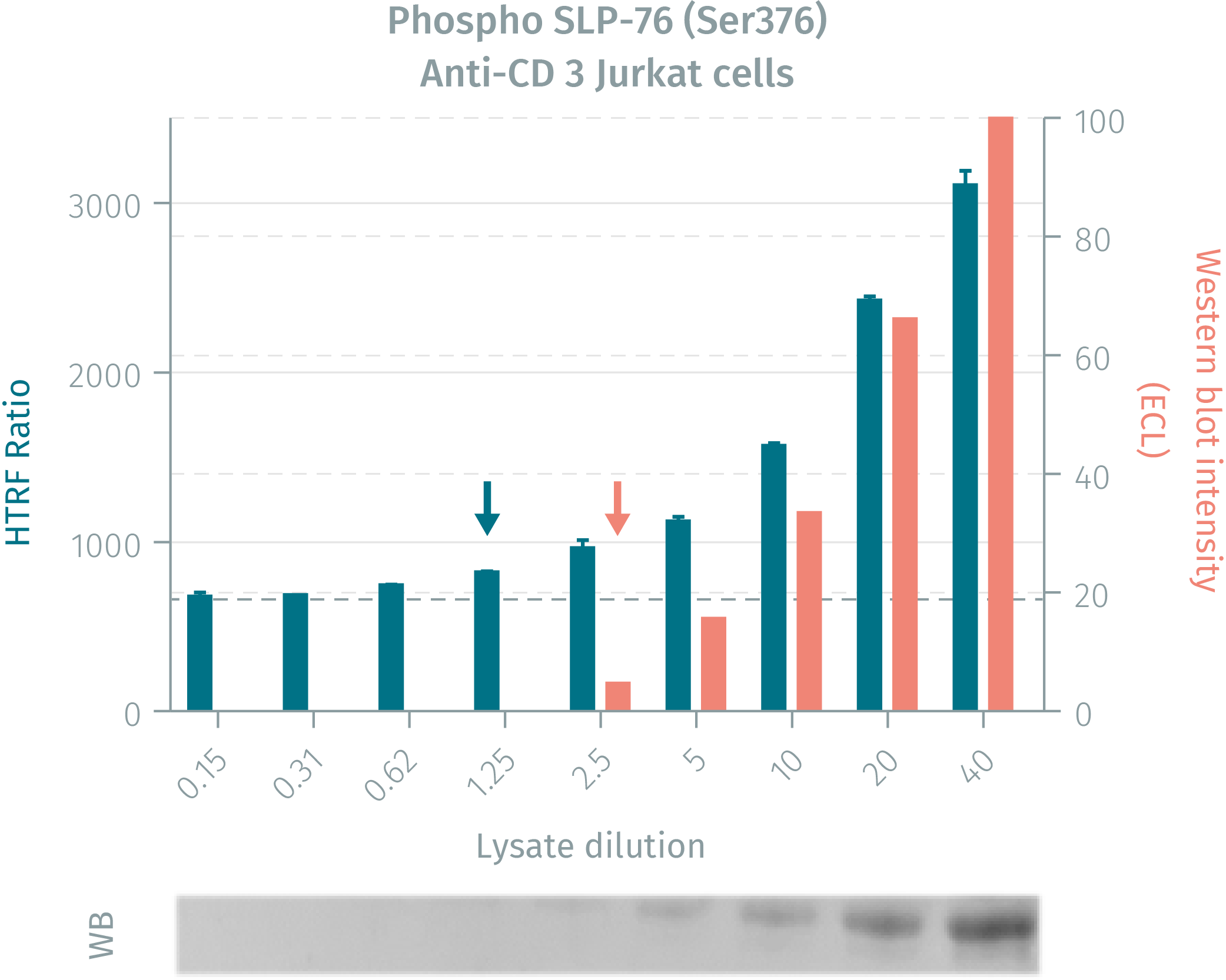
Assay validation
HTRF assay compared to western blot using phospho SLP-76 (Ser376) cellular assays on human jurkat cells
Jurkat cells were grown in a T175cm2 flask for 2 days in RPMI culture medium supplemented by 10% FBS, at 37°C in 5% CO2 atmosphere. 4.5 mL of cell suspension (13 x 106 cells / ml) were stimulated with anti-CD3 antibody (20µg/mL final concentration during 2.5min). After stimulation, the cells were lysed at RT for 30 min by adding 2.25 mL of 4X supplemented lysis buffer. Neat lysate was then serially diluted in the same supplemented lysis buffer. 16 µL of each dilution were analyzed in parallel by HTRF or by Western Blot.
Cell density optimisation for phospho SLP-76 (Ser376)
This cell density optimization was carried out using the two plate protocol. Jurkat cells were plated in a 96-well culture plate at 200,000 Kcells or 400Kcells in 25µL/well, in complete RPMI culture medium with 10% FBS. Cell stimulation was done by anti-CD3 Ab at various concentrations for 30 min. Cell lysis was performed using 10 µL of 4X supplemented lysis buffer. 16 µL of each cell lysate were analyzed by HTRF Phospho SLP-76(Ser376) assays

Kinetics of cell stimulation
This assay was carried out with a two plate protocol. 400,000 Jurkat cells per well were plated in a 96-well culture plate at 25µL/well in complete RPMI culture medium with 10% FBS. Cell stimulation was performed using anti CD3 Ab at the final concentration of 20µg/mL for different stimulation times (min). After stimulation, cells were lysed with 10 µL of 4X supplemented lysis buffer. 16 µL of each cell lysate for the different conditions were analyzed by HTRF Phospho SLP-76(Ser376) and Total SLP-76.

Monitoring of the immune checkpoint inhibitor PD-L1 activity
As Phospho SLP-76 (Tyr376) is a readout of T cell activation, it was used to monitor the inhibitory effect of PD-L1 recombinant protein on T cell activation. This assay was performed using the two-plate assay protocol. 400 000 Jurkat cells were plated in 25 µL of complete RPMI culture medium with 10% FBS. Cells were pre-incubated 24 hours with different concentrations of PD-L1 recombinant protein. Cells were then stimulated for 2.5 min with 20 µg / mL anti-CD3 Ab (final concentration) and lysed directly with10 µL of 4X supplemented lysis buffer, then incubated 30 min at RT. 16µL of each sample were analyzed using Phospho SLP-76 (Ser376) and Total SLP-76 assays. Phospho SLP-76 results were normalized with the Total SLP-76 amount and plotted on a graph.
A slight but significant inhibitory effect of PD-L1 recombinant protein was recorded with the Phospho SLP-76 (Ser376) assay at the final concentration of 40 µg/mL

Simplified pathway
Function and regulation of SLP-76
TCR engagement promotes the phosphorylation of immunoreceptor tyrosine-based activation motifs (ITAMs) on the cytosolic side of the TCR/CD3 complex by lymphocyte protein tyrosine kinase (Lck). Zap-70 is recruited to the TCR/CD3 complex, where it becomes phosphorylated and activated. ZAP-70 phosphorylates SLP-76. Activated SLP-76 translocates to the plasma membrane and promotes a multi-protein complex, which participates in the activation, survival, and proliferation of T-Lymphocytes

Resources
Are you looking for resources, click on the resource type to explore further.
Guide
HTRF solutions, guide to major applications
This guide provides you an overview of HTRF applications in several therapeutic areas.
Flyer
Reagent solutions for autoimmunity research.
Advance your autoimmune disease research and benefit from Revvity broad offering of reagent technologies
Loading...


How can we help you?
We are here to answer your questions.

