


US
Revvity Sites Globally
Select your location.
*e-commerce not available for this region.

HTRF Human & Mouse Phospho-S6RP (Ser235/236) Detection Kit, 10,000 Assay Points


HTRF Human & Mouse Phospho-S6RP (Ser235/236) Detection Kit, 10,000 Assay Points


Shipping box for Revvity reagent kits


S6rp Phospho-s235/236 Kit - 500 Tests
For research use only. Not for use in diagnostic procedures. All products to be used in accordance with applicable laws and regulations including without limitation, consumption and disposal requirements under European REACH regulations (EC 1907/2006).
| Feature | Specification |
|---|---|
| Application | Cell Signaling |
| Sample Volume | 16 µL |
S6rp Phospho-s235/236 Kit - 500 Tests
For research use only. Not for use in diagnostic procedures. All products to be used in accordance with applicable laws and regulations including without limitation, consumption and disposal requirements under European REACH regulations (EC 1907/2006).
Product Variants
Unit Size: 500 Assay Points
Part #:
64RP6PEG
List Price
USD 2,147.00
Unit Size: 10,000 Assay Points
Part #:
64RP6PEH
List Price
USD 12,490.00
HTRF Human & Mouse Phospho-S6RP (Ser235/236) Detection Kit, 10,000 Assay Points
Shipping box for Revvity reagent kits
HTRF Human & Mouse Phospho-S6RP (Ser235/236) Detection Kit, 10,000 Assay Points
Product information
Overview
The Phospho-S6RP (Ser235/236) cellular assay kit enables the quantitative detection of phosphorylated S6 ribosomal protein when phosphorylated on Serine 235/236. It can be used for the screening of receptor activation modulators and compounds acting on upstream events. This kit has various applications, from inflammation/immunology to metabolism/diabetes and oncology research.
Specifications
| Application |
Cell Signaling
|
|---|---|
| Brand |
HTRF
|
| Detection Modality |
HTRF
|
| Lysis Buffer Compatibility |
Lysis Buffer 1
Lysis Buffer 3
Lysis Buffer 4
Lysis Buffer 5
|
| Molecular Modification |
Phosphorylation
|
| Product Group |
Kit
|
| Sample Volume |
16 µL
|
| Shipping Conditions |
Shipped in Dry Ice
|
| Target Class |
Phosphoproteins
|
| Target Species |
Human
Mouse
|
| Technology |
TR-FRET
|
| Therapeutic Area |
Metabolism/Diabetes
Oncology & Inflammation
|
| Unit Size |
10,000 Assay Points
|
Video gallery
HTRF Human & Mouse Phospho-S6RP (Ser235/236) Detection Kit, 10,000 Assay Points
HTRF Human & Mouse Phospho-S6RP (Ser235/236) Detection Kit, 10,000 Assay Points
How it works
Phospho-S6RP (Ser235/236) assay principle
The Phospho-S6RP (Ser235/236) assay measures S6RP when phosphorylated at Ser235/236. Contrary to Western Blot, the assay is entirely plate-based and does not require gels, electrophoresis or transfer. The Phospho-S6RP (Ser235/236) assay uses 2 labeled antibodies: one with a donor fluorophore, the other one with an acceptor. The first antibody is selected for its specific binding to the phosphorylated motif on the protein, the second for its ability to recognize the protein independent of its phosphorylation state. Protein phosphorylation enables an immune-complex formation involving both labeled antibodies and which brings the donor fluorophore into close proximity to the acceptor, thereby generating a FRET signal. Its intensity is directly proportional to the concentration of phosphorylated protein present in the sample, and provides a means of assessing the protein's phosphorylation state under a no-wash assay format.
Phospho-S6RP (Ser235/236) 2-plate assay protocol
The 2 plate protocol involves culturing cells in a 96-well plate before lysis then transferring lysates to a 384-well low volume detection plate before adding phospho-S6RP (Ser235/236)HTRF detection reagents. This protocol enables the cells' viability and confluence to be monitored.

Phospho-S6RP (Ser235/236) 1-plate assay protocol
Detection of phosphorylated p-S6RP with HTRF reagents can be performed in a single plate used for culturing, stimulation and lysis. No washing steps are required. This HTS designed protocol enables miniaturization while maintaining robust HTRF quality.

Assay validation
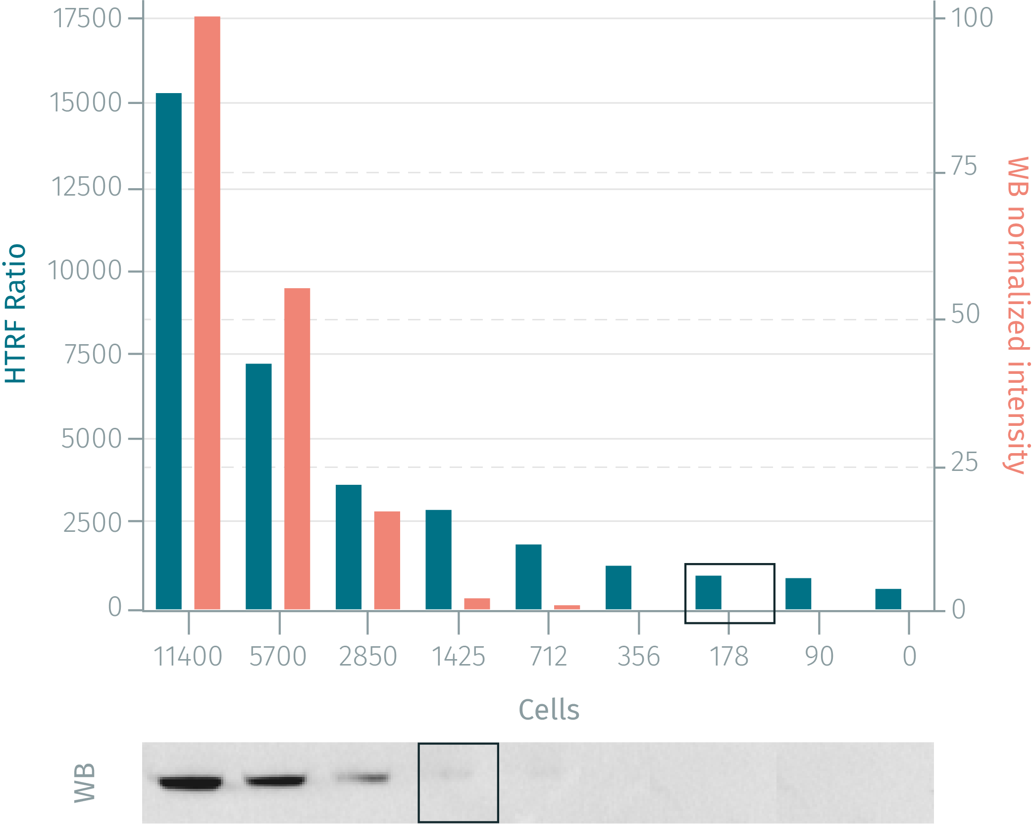
HTRF compared to WB using phospho-S6RP (Ser235/236) cellular assay kit
HeLa cells were grown in a T175 flask at 37°C, 5% CO2 for 1 day. After removal of cell culture medium, 3 mL of supplemented lysis buffer were added and incubated for 30 min. Soluble supernatants were collected after 10 min centrifuging. Equal amounts of lysates were used for a side by side comparison of WB and HTRF. HTRF phospho-S6RP assay shows better sensitivity than the Western Blot method.
Rapamycin dose response on murine NIH-3T3 or HEK293 cells
Murine NIH-3T3 or Human HEK293 cells were seeded in a cell culture-treated 96-well plate at various densities (from 12,500 to 100,000 cells/well). After an overnight serum starvation, increasing concentrations of rapamycin were applied from 2H, followed by a 20% serum stimulation for 30min. After a 30 minutes lysis incubation time with 200µL of supplemented lysis buffer, phosphorylated S6RP was measured using the two-plate Phospho-S6RP assay kit protocol. Note: depending on the cell lines used, lysis volume must be optimized (usually from 50µL to 200µL).

Simplified pathway
Phospho-S6RP simplified pathway
Ribosomes catalyze protein synthesis and are composed of a small 40S subunit and a large 60S subunit. The cytoplasmic S6 Ribosomal Protein (S6RP) is a component of the 40S subunit. RPS6 is the major substrate of protein kinases in the ribosome. Phosphorylation is induced by a wide range of stimuli, including growth factors, tumor-promoting agents, and mitogens. Dephosphorylation occurs at growth arrest. S6RP protein may contribute to the control of cell growth and proliferation through the selective translation of particular classes of mRNA.

Resources
Are you looking for resources, click on the resource type to explore further.
Guide
HTRF solutions, guide to major applications
This guide provides you an overview of HTRF applications in several therapeutic areas.


How can we help you?
We are here to answer your questions.

