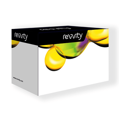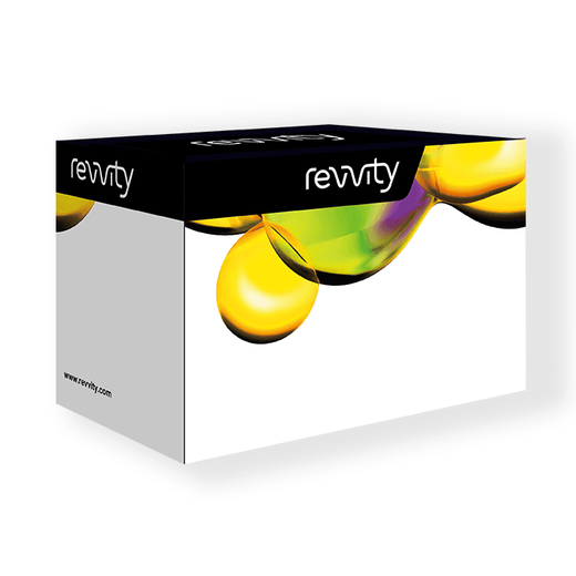

HTRF Total NDRG1 Detection Kit, 500 Assay Points


HTRF Total NDRG1 Detection Kit, 500 Assay Points


The total NDRG1 kit monitors the cellular level of NDRG1 and can be used as a normalization assay for the phospho-NDRG1 kits.
For research use only. Not for use in diagnostic procedures. All products to be used in accordance with applicable laws and regulations including without limitation, consumption and disposal requirements under European REACH regulations (EC 1907/2006).
| Feature | Specification |
|---|---|
| Application | Cell Signaling |
| Sample Volume | 16 µL |
The total NDRG1 kit monitors the cellular level of NDRG1 and can be used as a normalization assay for the phospho-NDRG1 kits.


HTRF Total NDRG1 Detection Kit, 500 Assay Points


HTRF Total NDRG1 Detection Kit, 500 Assay Points


Product information
Overview
The total NDRG1 kit serves as a normalization assay with HTRF Phospho-NDRG assay kits. NDRG1 is upregulated by HIF1-alpha under hypoxic conditions often associated with aggressive tumor progression. NDRG1 is a metastasis suppressor that works by blocking the angiogenesis signaling pathway.
Specifications
| Application |
Cell Signaling
|
|---|---|
| Brand |
HTRF
|
| Detection Modality |
HTRF
|
| Molecular Modification |
Total
|
| Product Group |
Kit
|
| Sample Volume |
16 µL
|
| Shipping Conditions |
Shipped in Dry Ice
|
| Target Class |
Phosphoproteins
|
| Technology |
TR-FRET
|
| Therapeutic Area |
Oncology & Inflammation
|
| Unit Size |
500 Assay Points
|
Video gallery

HTRF Total NDRG1 Detection Kit, 500 Assay Points

HTRF Total NDRG1 Detection Kit, 500 Assay Points

How it works
Total-NDRG1 assay principle
The Total-NDRG1 assay quantifies the expression level of NDRG1 in a cell lysate. Contrary to Western Blot, the assay is entirely plate-based and does not require gels, electrophoresis or transfer. The Total-NDRG1 assay uses two labeled antibodies: one coupled to a donor fluorophore, the other to an acceptor. Both antibodies are highly specific for a distinct epitope on the protein. In presence of NDRG1 in a cell extract, the addition of these conjugates brings the donor fluorophore into close proximity with the acceptor and thereby generates a FRET signal. Its intensity is directly proportional to the concentration of the protein present in the sample, and provides a means of assessing the proteins expression under a no-wash assay format.

Total-NDRG1 2-plate assay protocol
The 2 plate protocol involves culturing cells in a 96-well plate before lysis then transferring lysates to a 384-well low volume detection plate before adding Total NDRG1 HTRF detection reagents. This protocol enables the cells' viability and confluence to be monitored.

Total-NDRG1 1-plate assay protocol
Detection of total NDRG1 with HTRF reagents can be performed in a single plate used for culturing, stimulation and lysis. No washing steps are required. This HTS designed protocol enables miniaturization while maintaining robust HTRF quality.

Assay validation
Detection of phospho and total NDRG1 on HeLa cells
Different cell densities of HeLa cells were plated in 96-well plates and incubated overnight. Cells were stimulated with increasing concentration of 25 µL of AZD8055 (a novel ATP-competitive mTOR inhibitor) for 2h at 37°C - 5% CO2. After stimulation, cells were lysed with 25µL of lysis buffer 4X for 30 min at RT under gentle shaking. 16 µL of lysate were transferred into a 384-well sv white microplate and 4 µL of the HTRF phospho NDRG1 (Thr346) or total NDRG1 detection reagents were added. The HTRF signal was recorded after a overnight incubation at room temperature.


Detection of total NDRG1 on HeLa cells
Different cell densities (100K, 50K, 25K and 12.5K) of HeLa cells were plated under 50µL in 96-well plate in presence of 40 µM of Dp44mT and incubated for 24h. Then, media was aspirated and cells were lysed with 50µL of lysis buffer 1X for 30 min at RT under gentle shaking. 16 µL of lysate were transferred into a 384-well sv white microplate and 4 µL of the HTRF total NDRG1 detection reagents were added. The HTRF signal was recorded after a overnight incubation at room temperature.

Resources
Are you looking for resources, click on the resource type to explore further.
This guide provides you an overview of HTRF applications in several therapeutic areas.
SDS, COAs, Manuals and more
Are you looking for technical documents related to the product? We have categorized them in dedicated sections below. Explore now.
-
LanguageEnglishCountryUnited States
-
LanguageFrenchCountryFrance
-
LanguageGermanCountryGermany
-
LanguageGreekCountryGreece
-
LanguageItalianCountryItaly
-
LanguageSpanishCountrySpain
-
LanguageEnglishCountryUnited Kingdom
-
Resource typeManualLanguageEnglishCountry-


How can we help you?
We are here to answer your questions.


