


US
Revvity Sites Globally
Select your location.
*e-commerce not available for this region.

HTRF Human & Mouse Phospho-MEK1 (Ser218/222) Detection Kit, 500 Assay Points


HTRF Human & Mouse Phospho-MEK1 (Ser218/222) Detection Kit, 500 Assay Points


Shipping box for Revvity reagent kits


The Phospho-MEK1 (Ser218/222) kit enables the cell-based detection of MEK1 protein phosphorylated at Ser218/222 when studying the RAS-RAF-MEK cascade in the MAPK/ERK pathway.
For research use only. Not for use in diagnostic procedures. All products to be used in accordance with applicable laws and regulations including without limitation, consumption and disposal requirements under European REACH regulations (EC 1907/2006).
| Feature | Specification |
|---|---|
| Application | Cell Signaling |
| Sample Volume | 16 µL |
The Phospho-MEK1 (Ser218/222) kit enables the cell-based detection of MEK1 protein phosphorylated at Ser218/222 when studying the RAS-RAF-MEK cascade in the MAPK/ERK pathway.
For research use only. Not for use in diagnostic procedures. All products to be used in accordance with applicable laws and regulations including without limitation, consumption and disposal requirements under European REACH regulations (EC 1907/2006).
Product Variants
Unit Size: 500 Assay Points
Part #:
64ME1PEG
List Price
USD 2,147.00
Unit Size: 10,000 Assay Points
Part #:
64ME1PEH
List Price
USD 12,490.00
HTRF Human & Mouse Phospho-MEK1 (Ser218/222) Detection Kit, 500 Assay Points
Shipping box for Revvity reagent kits
HTRF Human & Mouse Phospho-MEK1 (Ser218/222) Detection Kit, 500 Assay Points
Product information
Overview
The Phospho-MEK1 (Ser218/222) kit measures endogenous MEK1 protein phosphorylated at Ser218/222 when studying the RAS-RAF-MEK cascade in the MAPK/ERK pathway. Phosphorylated by RAF, this dual specificity mitogen-activated protein kinase is responsible for the phosphorylation of ERK, one of the most important signaling nodes, and protein kinase in cell proliferation, survival, and differentiation pathways.
Specifications
| Application |
Cell Signaling
|
|---|---|
| Brand |
HTRF
|
| Detection Modality |
HTRF
|
| Lysis Buffer Compatibility |
Lysis Buffer 1
|
| Molecular Modification |
Phosphorylation
|
| Product Group |
Kit
|
| Sample Volume |
16 µL
|
| Shipping Conditions |
Shipped in Dry Ice
|
| Target Class |
Phosphoproteins
|
| Target Species |
Human
Mouse
|
| Technology |
TR-FRET
|
| Therapeutic Area |
Cardiovascular
Neuroscience
Oncology & Inflammation
|
| Unit Size |
500 Assay Points
|
Video gallery
HTRF Human & Mouse Phospho-MEK1 (Ser218/222) Detection Kit, 500 Assay Points
HTRF Human & Mouse Phospho-MEK1 (Ser218/222) Detection Kit, 500 Assay Points
How it works
Phospho-MEK1 (Ser218/222) assay principle
The Phospho-MEK1 (Ser218/222) assay measures MEK1 when phosphorylated at Ser218/222. Contrary to Western Blot, the assay is entirely plate-based and does not require gels, electrophoresis or transfer. The Phospho-MEK1 (Ser218/222) assay uses 2 labeled antibodies: one with a donor fluorophore, the other one with an acceptor. The first antibody is selected for its specific binding to the phosphorylated motif on the protein, the second for its ability to recognize the protein independent of its phosphorylation state. Protein phosphorylation enables an immune-complex formation involving both labeled antibodies and which brings the donor fluorophore into close proximity to the acceptor, thereby generating a FRET signal. Its intensity is directly proportional to the concentration of phosphorylated protein present in the sample, and provides a means of assessing the proteins phosphorylation state under a no-wash assay format.
Phospho-MEK1 (Ser218/222) 2-plate assay protocol
The 2 plate protocol involves culturing cells in a 96-well plate before lysis then transferring lysates to a 384-well low volume detection plate before adding Phospho-MEK1 (Ser218/222) HTRF detection reagents. This protocol enables the cells' viability and confluence to be monitored.

Phospho-MEK1 (Ser218/222) 1-plate assay protocol
Detection of Phosphorylated MEK1 (Ser218/222) with HTRF reagents can be performed in a single plate used for culturing, stimulation and lysis. No washing steps are required. This HTS designed protocol enables miniaturization while maintaining robust HTRF quality.

Assay validation
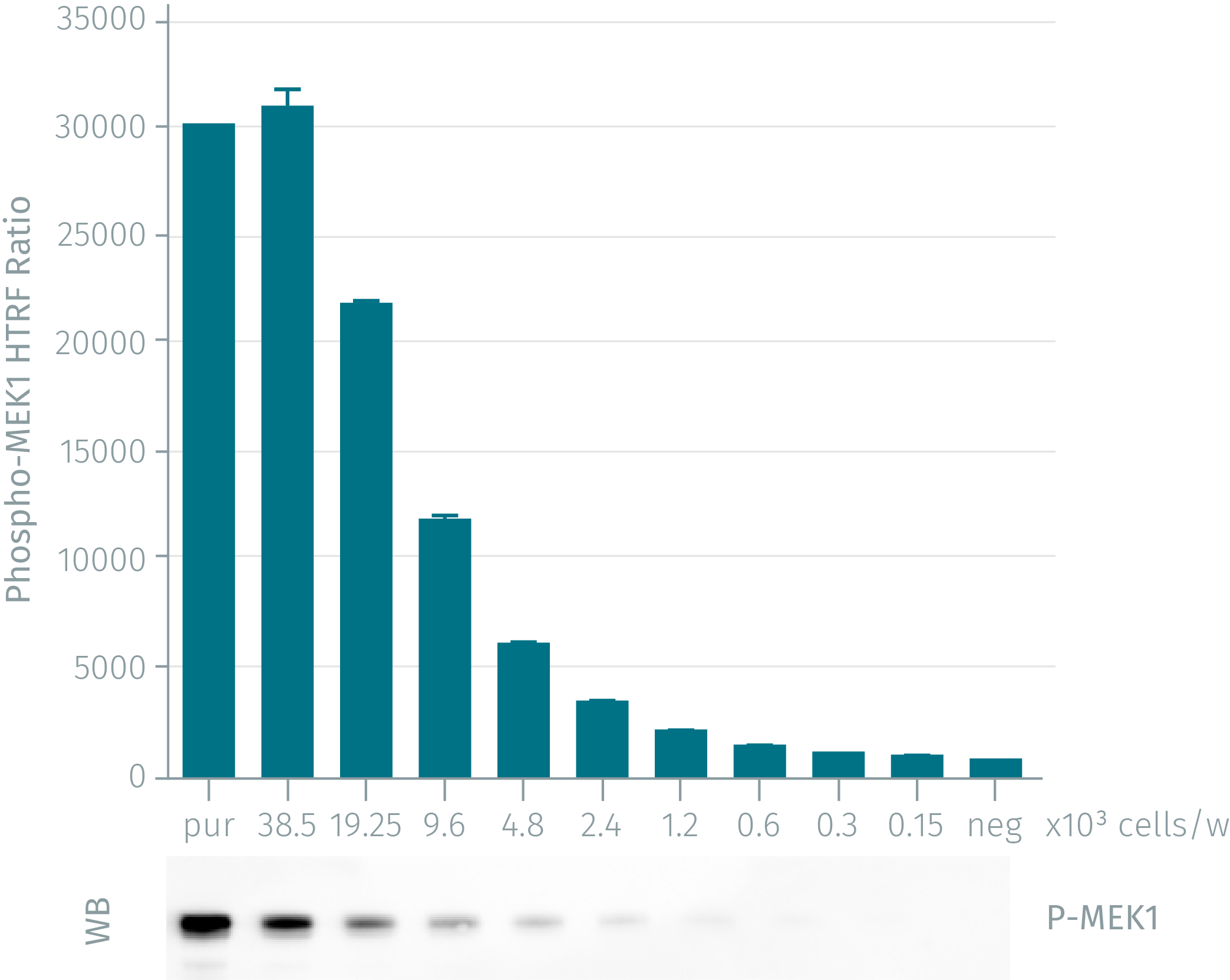
WB versus HTRF assay for phospho-MEK1 (Ser218/222)
HeLa cells were grown in a T175 flask 37°C, 5% Co2, 2 days. Stimulation was done with EGF 2nM for 5 min. After removal of cell culture medium, 3 mL of supplemented lysis buffer were added and incubated for 30 min. Soluble supernatants were collected after 10 min centrifuging. Equal amounts of lysates were used for a side by side comparison of WB and HTRF. Phospho-MEK1 assay shows the same level of sensitivity as Western Blot.
PMA dose-response on NIH 3T3 cells
Murine NIH 3T3 cells (25,000 cells/well) were stimulated for 15 minutes at 37°C with various concentrations of PMA. After a 30 minutess lysis incubation time, phosphorylated MEK1 were measured using the two-plate assay protocol.

Inhibition effect of EGRF & RAF inhibitors on HeLa cells
HeLa cells (50,000 cells/well) were incubated for 15 min at 37°C with various concentrations of the 2 inhibitors, Erlotinib (the EGFR inhibitor) and L777,450 (a Raf inhibitor). EGF 0.8nM (agonist) was then added and incubated for 10 minutes. After a 30 minutes lysis incubation time, inhibition of the phosphorylated MEK was measured using the Phospho-MEK1 assay. Assay was done using the two-plate assay protocol.

Resources
Are you looking for resources, click on the resource type to explore further.
Application Note
A guideline for HTRF cell-based phospho-protein data normalization
Get the best out of your phosphorylation assays
Combining phospho and total protein assays enables better analysis. This...
Application Note
Detection of MAPK activation to evaluate the efficacy and potency of KRAS/SOS1 inhibitors by AlphaLISA and HTRF technologies
Evaluation of the therapeutic profile of anti-oncogene compounds in various cell lines with AlphaLISA™ and HTRF™
KRAS is a proto...
Brochure
HTRF assays and reagents catalog
Discover the versatility and precision of Homogeneous Time-Resolved Fluorescence (HTRF) technology. Our HTRF portfolio offers a...
Guide
HTRF solutions, guide to major applications
This guide provides you an overview of HTRF applications in several therapeutic areas.
Brochure
Signaling in the immune system - HTRF assays for oncology and inflammation
Download this summary of signaling pathways in the immune system with HTRF targets.
Included:
- Detailed pathways about NK cells, T...
Guide
Understanding obesity: exploring cellular pathways and mechanisms
Obesity is a complex condition characterized by excessive fat accumulation, posing significant health and socioeconomic challenges...


How can we help you?
We are here to answer your questions.

