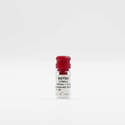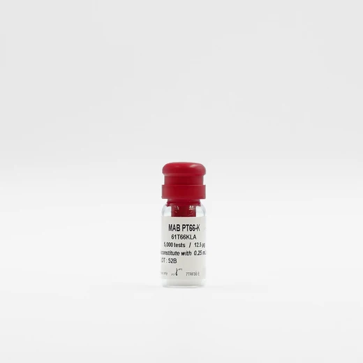

HTRF MAb PT66-Eu cryptate, 5,000 Assay Points


HTRF MAb PT66-Eu cryptate, 5,000 Assay Points


MAb PT66-Eu cryptate enables the rapid detection of phosphorylation of peptides or proteins on Tyrosine in a biochemical kinase assay format.
For research use only. Not for use in diagnostic procedures.
| Feature | Specification |
|---|---|
| Application | Biochemical Enzymatic Assay |
MAb PT66-Eu cryptate enables the rapid detection of phosphorylation of peptides or proteins on Tyrosine in a biochemical kinase assay format.
For research use only. Not for use in diagnostic procedures.


HTRF MAb PT66-Eu cryptate, 5,000 Assay Points


HTRF MAb PT66-Eu cryptate, 5,000 Assay Points


Product information
Overview
PT66-Eu cryptate is a labeled mouse monoclonal IgG1. It shows to react specifically with phosphorylated tyrosine, both as free amino acid or when conjugated to carriers. No cross-reactivity has been observed with non-phosphorylated tyrosine, phosphothreonine, phosphoserine, AMP or ATP. This allows the construction of a highly specific biochemical Tyrosine kinase assay. Any kind of substrate can be used, from small synthetic peptides to natural substrates, meaning high assay detection versatility. HTRF allows to perform assay at physiological ATP concentrations.
Specifications
| Application |
Biochemical Enzymatic Assay
|
|---|---|
| Brand |
HTRF
|
| Detection Modality |
HTRF
|
| Product Group |
Fluorescent Reagent
|
| Shipping Conditions |
Shipped Ambient
|
| Target Class |
Kinases
|
| Technology |
TR-FRET
|
| Therapeutic Area |
Cardiovascular
Infectious Diseases
Inflammation
Metabolism/Diabetes
NASH/Fibrosis
Neuroscience
Oncology & Inflammation
Rare Diseases
|
| Unit Size |
5,000 Assay Points
|
Video gallery

HTRF MAb PT66-Eu cryptate, 5,000 Assay Points

HTRF MAb PT66-Eu cryptate, 5,000 Assay Points

How it works
Assay principle
HTRF kinase assays typically use a combination of a Europium-cryptate labeled anti-phosphoresidue antibody, SA-XL665 and a biotinylated* substrate. The signal is proportional to the concentration of phospho-residues. *Alternatively, other tags such as 6HIS, GST, c-myc, DNP, or FLAG may be used to label the kinase substrate.

Assay protocol
HTRF kinase assays have two basic phases: 1. Enzymatic step: biotinylated substrate is incubated with the kinase of interest and with the compounds in presence of co-factors. The reaction starts with the addition of ATP. 2. Detection step: The phosphorylated biotinylated substrate is then detected by the addition of Streptavidin-XL665 and MAb PT66-Eu cryptate prepared in a buffer containing EDTA to stop the enzymatic reaction.

Assay validation
Detection at physiological ATP concentration
ATP concentration is an important factor for in vitro kinase assay design, as the sensitivity of ATP competitive inhibitors varies with ATP concentration. As shown in Figure 5, HTRF tolerates the extremely high ATP concentrations (up to mM range) needed for mechanistic studies, e.g. the confirmation of ATP competitive inhibitors. The shift observed for increasing ATP concentrations in the graph confirms the ATP-competitive effect of the compound tested. Assay was performed on a tyrosine kinase revealed by PT66.

Membrane friendly
In addition to fresh and frozen cells, the HTRF Kinase toolbox can be used with membrane preparations. The graph depicts an assay scheme for measuring autophosphorylation of a receptor tyrosine kinase (RTK), using membrane preparations as a source of active kinase. Autophosphorylation of an RTK expressed as a tagged fusion protein is detected using a Eu3+ labeled anti-phospho tyrosine antibody and XL665 labeled anti-tag antibody.

Resources
Are you looking for resources, click on the resource type to explore further.
This guide provides you an overview of HTRF applications in several therapeutic areas.


How can we help you?
We are here to answer your questions.


