


US
Revvity Sites Globally
Select your location.
*e-commerce not available for this region.

HTRF Phospho-IR-β (Insulin Receptor Beta) (Tyr1150 /1151) Detection Kit, 500 Assay Points
")

HTRF Phospho-IR-β (Insulin Receptor Beta) (Tyr1150 /1151) Detection Kit, 500 Assay Points
")

PHOTO phospho Insulin receptor Tyr1150/1151 kit
")

This HTRF assay enables the cell-based detection of Tyr1150/1151 phosphorylation on Insulin Receptor-beta.
For research use only. Not for use in diagnostic procedures. All products to be used in accordance with applicable laws and regulations including without limitation, consumption and disposal requirements under European REACH regulations (EC 1907/2006).
| Feature | Specification |
|---|---|
| Application | Cell Signaling |
| Sample Volume | 16 µL |
This HTRF assay enables the cell-based detection of Tyr1150/1151 phosphorylation on Insulin Receptor-beta.
For research use only. Not for use in diagnostic procedures. All products to be used in accordance with applicable laws and regulations including without limitation, consumption and disposal requirements under European REACH regulations (EC 1907/2006).
Product Variants
Unit Size: 500 Assay Points
Part #:
63ADK016PEG
List Price
USD 2,147.00
Unit Size: 10,000 Assay Points
Part #:
63ADK016PEH
List Price
USD 12,490.00
HTRF Phospho-IR-β (Insulin Receptor Beta) (Tyr1150 /1151) Detection Kit, 500 Assay Points
PHOTO phospho Insulin receptor Tyr1150/1151 kit
HTRF Phospho-IR-β (Insulin Receptor Beta) (Tyr1150 /1151) Detection Kit, 500 Assay Points
Product information
Overview
Phospho-Insulin Receptor-beta (Tyr1150/1151) assay enables the detection of Tyr1150/1151 phosphorylation on Insulin Receptor-beta. Stimulated by insulin and insulin-like growth factors, the insulin receptor is involved in several pathways such as those for Insulin and FoxO signaling.
Specifications
| Application |
Cell Signaling
|
|---|---|
| Brand |
HTRF
|
| Detection Modality |
HTRF
|
| Molecular Modification |
Phosphorylation
|
| Product Group |
Kit
|
| Sample Volume |
16 µL
|
| Shipping Conditions |
Shipped in Dry Ice
|
| Target Class |
Phosphoproteins
|
| Technology |
TR-FRET
|
| Therapeutic Area |
Metabolism/Diabetes
Neuroscience
|
| Unit Size |
500 Assay Points
|
Video gallery
HTRF Phospho-IR-β (Insulin Receptor Beta) (Tyr1150 /1151) Detection Kit, 500 Assay Points
HTRF Phospho-IR-β (Insulin Receptor Beta) (Tyr1150 /1151) Detection Kit, 500 Assay Points
How it works
Phospho-Insulin Receptor ß (Ser177/181) assay principle
The Phospho-Insulin Receptor-beta (Ser177/181) assay measures Insulin Receptor-beta when phosphorylated at Ser177/181. Contrary to Western Blot, the assay is entirely plate-based and does not require gels, electrophoresis or transfer. The Phospho-Insulin Receptor-beta (Ser177/181) assay uses 2 labeled antibodies: one with a donor fluorophore, the other one with an acceptor. The first antibody is selected for its specific binding to the phosphorylated motif on the protein, the second for its ability to recognize the protein independent of its phosphorylation state. Protein phosphorylation enables an immune-complex formation involving both labeled antibodies and which brings the donor fluorophore into close proximity to the acceptor, thereby generating a FRET signal. Its intensity is directly proportional to the concentration of phosphorylated protein present in the sample, and provides a means of assessing the protein's phosphorylation state under a no-wash assay format.
Phospho-Insulin Receptor ß (Ser177/181) 2-plate assay protocol
The 2 plate protocol involves culturing cells in a 96-well plate before lysis then transferring lysates to a 384-well low volume detection plate before adding Phospho-IR-beta (Ser177/181) HTRF detection reagents. This protocol enables the cells' viability and confluence to be monitored.

Phospho-Insulin Receptor ß (Ser177/181) 1-plate assay protocol
Detection of Phosphorylated IR-beta (Ser177/181) with HTRF reagents can be performed in a single plate used for culturing, stimulation and lysis. No washing steps are required. This HTS designed protocol enables miniaturization while maintaining robust HTRF quality.

Assay validation
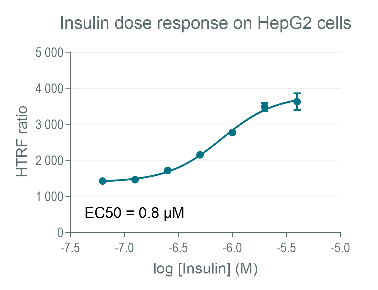
Phospho-IRβ (Tyr1150/1151) illustration on HepG2 cells
HepG2 cells were plated at 100,000 cells per well on a 96-well plate. After an overnight incubation at 37°C, 5% CO2, a serial dilution of insulin was added under cells for 5 minutes at 37°C, 5% CO2. Stimulation medium was removed from cells and 50µL of lysis buffer was added onto the cells. Lysis step was done by shaking gently during 30 minutes. 16µL of samples were transferred in a 384-well small volume plate then 4µL of Phospho IRβ HTRF detection reagents were added. Signals were recorded overnight.
Phospho-IRβ (Tyr1150/1151) illustration on HepaRG
HepaRG cells were plated at 200,000 cells per well on a 96-well plate. After an incubation of 5 days at 37°C, 5% CO2, a serial dilution of insulin was added under cells in presence of pervanadate at 30µM for 5 minutes at 37°C, 5% CO2. Stimulation medium was removed from cells and 50µL of lysis buffer was added onto the cells. Lysis step was done by shaking gently during 30 minutes. 16µL of samples were transferred in a 384-well small volume plate then 4µL of phospho IRβ HTRF detection reagents were added. In parallel 16µl were dispensed on other wells than 4µL of total IRβ HTRF detection reagents were added. Signals were recorded overnight.

Phospho-IRβ (Tyr1150/1151) WB comparison
Human Hek 293 cells were cultured to 80% confluency. After insulin and pervanadate co-treatment, cells were lysed and soluble supernatants were collected via centrifugation. Serial dilutions of the cell lysate were performed and 16 µL of each dilution were transferred into a 384-well low volume white microplate before finally adding phospho IRβ HTRF cellular kit reagents. A side by side comparison showed the HTRF Phospho assay is at least 8-fold more sensitive than the Western Blot.

Resources
Are you looking for resources, click on the resource type to explore further.
Guide
HTRF solutions, guide to major applications
This guide provides you an overview of HTRF applications in several therapeutic areas.
Guide
Understanding obesity: exploring cellular pathways and mechanisms
Obesity is a complex condition characterized by excessive fat accumulation, posing significant health and socioeconomic challenges...


How can we help you?
We are here to answer your questions.

