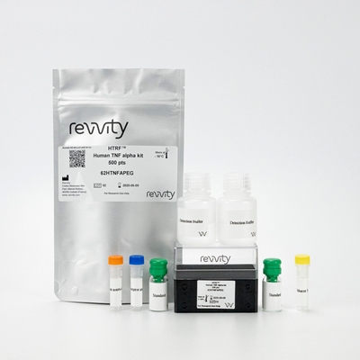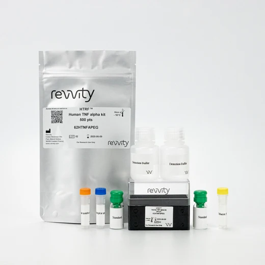

HTRF Human TNα Detection Kit, 10,000 Assay Points


HTRF Human TNα Detection Kit, 10,000 Assay Points


The HTRF human TNFα kit is designed for the quantification of human TNF alpha release in cell supernatant.
For research use only. Not for use in diagnostic procedures. All products to be used in accordance with applicable laws and regulations including without limitation, consumption and disposal requirements under European REACH regulations (EC 1907/2006).
| Feature | Specification |
|---|---|
| Application | Protein Quantification |
| Sample Volume | 16 µL |
The HTRF human TNFα kit is designed for the quantification of human TNF alpha release in cell supernatant.
For research use only. Not for use in diagnostic procedures. All products to be used in accordance with applicable laws and regulations including without limitation, consumption and disposal requirements under European REACH regulations (EC 1907/2006).


HTRF Human TNα Detection Kit, 10,000 Assay Points


HTRF Human TNα Detection Kit, 10,000 Assay Points


Product information
Overview
TNF alpha is a cytokine mainly produced by macrophages, T cells, and neutrophils. TNF alpha acts as a chemoattractant neutrophils, and promotes infiltration and activation at inflammation sites. TNF alpha is involved in several cancer types its ability to promote angiogenesis and cell proliferation, as well as to inhibit apoptosis.
Assessment of serum samples often requires enhanced sensitivity. In some cases, AlphaLISA assays may have sufficient sensitivity to enable detection of low levels of analytes in serum or plasma. When assaying, always follow recommended protocol and avoid highly haemolyzed samples.
Specifications
| Application |
Protein Quantification
|
|---|---|
| Brand |
HTRF
|
| Detection Modality |
HTRF
|
| Product Group |
Kit
|
| Sample Volume |
16 µL
|
| Shipping Conditions |
Shipped in Dry Ice
|
| Target Class |
Cytokines
|
| Target Species |
Human
|
| Technology |
TR-FRET
|
| Therapeutic Area |
Metabolism/Diabetes
NASH/Fibrosis
Oncology & Inflammation
|
| Unit Size |
10,000 Assay Points
|
Video gallery

HTRF Human TNα Detection Kit, 10,000 Assay Points

HTRF Human TNα Detection Kit, 10,000 Assay Points

Citations
How it works
Assay principle
Cell supernatant, sample, or standard is dispensed directly into the assay plate for the detection by HTRF® reagents (384-well low-volume white plate or Revvity low-volume 96-well plate in 20 µl). The antibodies labeled with the HTRF donor and acceptor are pre-mixed and added in a single dispensing step, to further streamline the assay procedure. The assay can be run up to a 1536-well format by simply resizing each addition volume proportionally.

Assay data analysis
The 4 Parameter Logistic (4PL) curve is commonly recommended for fitting an ELISA standard curve. This regression enables the accurate measurement of an unknown sample across a wider range of concentrations than linear analysis, making it ideally suited to the analysis of biological systems like cytokine releases.
To fully understand how to deal with HTRF data processing and also 4PL 1/y² fitting, Revvity also worked with Myassays.com to help you in your data analysis. you’ll be able to access a free online software to run your TNF alpha analysis.
Assay details
Technical specifications of human TNF alpha kit
| Sample size | 16 µL |
|---|---|
| Final assay volume | 20 µL |
| Kit components | Lyophilized standard, frozen detection antibodies, buffers &protocol. |
| LOD &LOQ (in Diluent) | 5 pg/mL &29 pg/mL |
| Range | 29 2,500 pg/mL |
| Time to result | 2h at RT |
| Calibration | NIBSC (12/154) value (IU/mL) = 0,1 x HTRF hTNFa value (pg/mL) |
| Species | Human only |
Analytical performance
Intra and inter assay
Intra-assay (n=24)
| Sample | Mean [TNFα] (pg/mL) | CV |
|---|---|---|
| 1 | 113 | 6% |
| 2 | 344 | 3% |
| 3 | 1399 | 6% |
| Mean CV | 5% |
Each of the 3 samples was measured 24 times, and % CV was calculated for each sample.
Inter-assay (n=4)
| Sample | [TNFa] (pg/mL) | Mean (delta R) | CV |
|---|---|---|---|
| 1 | 78 | 272 | 11% |
| 2 | 312 | 911 | 7% |
| 3 | 1250 | 2894 | 6% |
| Mean CV | 6% |
Each of the samples was measured in 4 different experiments, and % CV was calculated for each sample.
Dilutional linearity
The excellent % of recovery obtained from these experiments show the good linearity of the assay.
|
Sample |
Dilution factor | [TNFα] expected (pg/mL) | [TNFα] detected (pg/mL) | Recovery |
|---|---|---|---|---|
| 1 | 1 | - | 2336 | - |
| 3 | 782 | 898 | 115% | |
| 4.3 | 543 | 567 | 104% | |
| 10.7 | 218 | 252 | 116% | |
| Mean | 112% | |||
| 2 | 1 | - | 449 | - |
| 3 | 150 | 179 | 119% | |
| Mean | 119% | |||
Spike and recovery
|
Sample |
[TNFα] added (pg/mL) | [TNFα] expected (pg/mL) | [TNFα] detected (pg/mL) | Recovery |
|---|---|---|---|---|
| 1 | 1,250 | 1,555 | 1,523 | 98% |
| 2 | 1,250 | 1,874 | 1,892 | 101% |
Assay validation
Inhibition of TNF alpha secretion in THP1 cells with JTE607
THP1 cells plated at 100 kcells/well were incubated with increasing concentrations of JTE 607 for 18h, then stimulated for 3 h with 2 µg/mL LPS. 16 µL of supernatants were then transferred into a white detection plate (384 low volume) to be analyzed by the Human TNFa Assay.

TNF alpha secretion in PBMCs stimulated with LPS
PBMC plated at 50, 100, 200 and 400 kcells/well were stimulated for 3h with increasing concentrations of LPS (0, 0.02, 0.2, 2 µg/mL). 16 µL of supernatants were then transferred into a white detection plate (384 low volume) to be analyzed by the Human TNFa Assay.

Resources
Are you looking for resources, click on the resource type to explore further.
This application note demonstrates how compound’s mechanism of action and potential cytotoxic effects can be deciphered thanks to...
SDS, COAs, Manuals and more
Are you looking for technical documents related to the product? We have categorized them in dedicated sections below. Explore now.
-
LanguageEnglishCountryUnited States
-
LanguageFrenchCountryFrance
-
LanguageGermanCountryGermany
-
LanguageGreekCountryGreece
-
LanguageItalianCountryItaly
-
LanguageSpanishCountrySpain
-
LanguageEnglishCountryUnited Kingdom
-
Lot number11ALot dateOctober 2, 2026
-
Lot number10CLot dateMarch 15, 2026
-
Lot number12ALot dateSeptember 7, 2025
-
Lot number10BLot dateSeptember 7, 2025
-
Resource typeManualLanguageEnglishCountry-


How can we help you?
We are here to answer your questions.


