


US
Revvity Sites Globally
Select your location.
*e-commerce not available for this region.

HTRF Human & Mouse Phospho-Histone H3 (Thr3) Detection Kit, 500 Assay Points
")

HTRF Human & Mouse Phospho-Histone H3 (Thr3) Detection Kit, 500 Assay Points
")

PHOTO Phospho Histone H3 Thr3 kit
")

The phospho-Histone H3 (Thr3) assay enables the cell-based detection of Thr3 phosphorylation of Histone H3, a key regulator of gene expression and mitosis.
For research use only. Not for use in diagnostic procedures. All products to be used in accordance with applicable laws and regulations including without limitation, consumption and disposal requirements under European REACH regulations (EC 1907/2006).
| Feature | Specification |
|---|---|
| Application | Cell Signaling |
| Sample Volume | 16 µL |
The phospho-Histone H3 (Thr3) assay enables the cell-based detection of Thr3 phosphorylation of Histone H3, a key regulator of gene expression and mitosis.
For research use only. Not for use in diagnostic procedures. All products to be used in accordance with applicable laws and regulations including without limitation, consumption and disposal requirements under European REACH regulations (EC 1907/2006).
Product Variants
Unit Size: 500 Assay Points
Part #:
63ADK061PEG
List Price
USD 2,147.00
Unit Size: 10,000 Assay Points
Part #:
63ADK061PEH
List Price
USD 12,490.00
HTRF Human & Mouse Phospho-Histone H3 (Thr3) Detection Kit, 500 Assay Points
PHOTO Phospho Histone H3 Thr3 kit
HTRF Human & Mouse Phospho-Histone H3 (Thr3) Detection Kit, 500 Assay Points
Product information
Overview
Phospho-Histone H3 (T3) assay enables the detection of T3 phosphorylation in the tails of Histone H3, a key regulator of gene expression and mitosis. Phosphorylation at Threonine 3, catalyzed by the kinase haspin and highly conserved among many species, leads to recruitment of Aurora kinase B (AURKB) and the chromosomal passenger complex (CPC) to kinetochores, to regulate mitosis.
Specifications
| Application |
Cell Signaling
|
|---|---|
| Brand |
HTRF
|
| Detection Modality |
HTRF
|
| Lysis Buffer Compatibility |
Lysis Buffer 1
|
| Molecular Modification |
Phosphorylation
|
| Product Group |
Kit
|
| Sample Volume |
16 µL
|
| Shipping Conditions |
Shipped in Dry Ice
|
| Target Class |
Phosphoproteins
|
| Target Species |
Human
Mouse
|
| Technology |
TR-FRET
|
| Therapeutic Area |
Neuroscience
|
| Unit Size |
500 Assay Points
|
Video gallery
HTRF Human & Mouse Phospho-Histone H3 (Thr3) Detection Kit, 500 Assay Points
HTRF Human & Mouse Phospho-Histone H3 (Thr3) Detection Kit, 500 Assay Points
How it works
Phospho-Histone H3 (Thr3) assay principle
The phospho-Histone H3 (Thr3) assay measures Histone H3 when phosphorylated at Thr3. Contrary to Western Blot, the assay is entirely plate-based and does not require gels, electrophoresis or transfer. The phospho-Histone H3 (Thr3) assay uses 2 labeled antibodies: one with a donor fluorophore, the other one with an acceptor. The first antibody is selected for its specific binding to the phosphorylated motif on the protein, the second for its ability to recognize the protein independent of its phosphorylation state. Protein phosphorylation enables an immune-complex formation involving both labeled antibodies and which brings the donor fluorophore into close proximity to the acceptor, thereby generating a FRET signal. Its intensity is directly proportional to the concentration of phosphorylated protein present in the sample, and provides a means of assessing the protein’s phosphorylation state under a no-wash assay format.
Phospho-Histone H3 (Thr3) 2-plate assay protocol
The 2 plate protocol involves culturing cells in a 96-well plate before lysis then transferring lysates to a 384-well low volume detection plate before adding phospho-Histone H3 (Thr3) HTRF detection reagents. This protocol enables the cells' viability and confluence to be monitored.

Phospho-Histone H3 (Thr3) 1-plate assay protocol
Detection of Phosphorylated Histone H3 (Thr3) with HTRF reagents can be performed in a single plate used for culturing, stimulation and lysis. No washing steps are required. This HTS designed protocol enables miniaturization while maintaining robust HTRF quality.

Assay validation
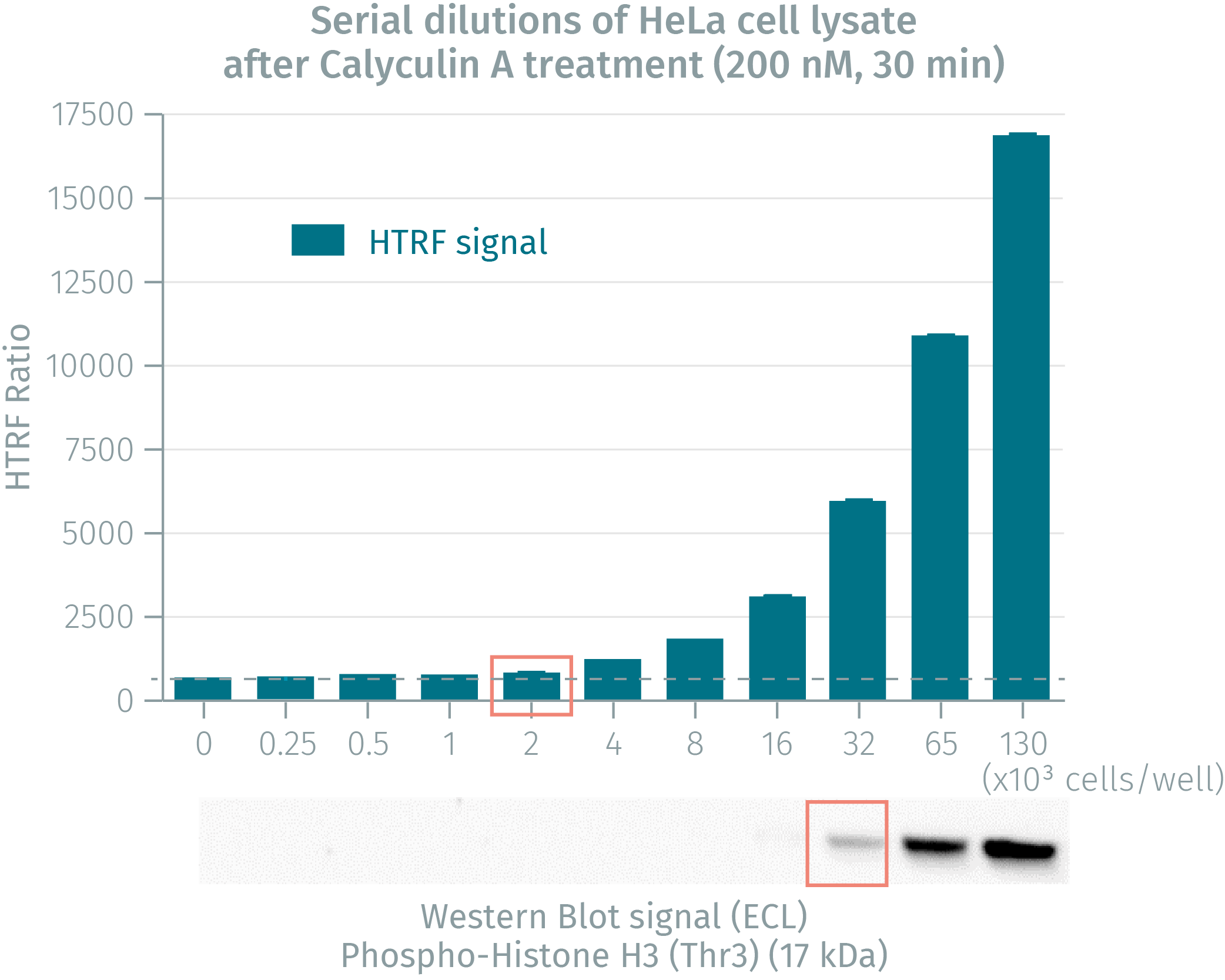
HTRF phospho H3 (Thr3) assay vs western blot
Human cervical cancer HeLa cells were cultured until reaching 80% confluency. After treatment with Calyculin A, cells were lysed and soluble fractions collected after a 10 min centrifugation. Serial dilutions of the cell lysate were performed, dispensed and analyzed side-by-side by Western Blot and by HTRF. The HTRF phospho H3 (Thr3) assay is at least 16-fold more sensitive than Western Blot.
HTRF phospho H3 (Thr3) validation assay on mouse and human cells
50,000 human cervical cancer HeLa cells or mouse fibroblast NIH-3T3 cells were plated in 96-well plates and incubated for 24h at 37 °C - 5% CO2. Cells were stimulated at 37°C for 30 min with increasing concentration of Calyculin-A. Medium was then removed and cells were lysed with 50µl of lysis buffer for 30min at RT under gentle shaking. 16 µL of lysate were transferred into a 384-well sv white microplate and 4 µL of the HTRF phospho-Histone H3 (Thr3) detection reagents were added. The HTRF signal was recorded after a 2h incubation time at room temperature.


Simplified pathway
Phospho-Histone H3-Thr3 in cell signaling
There are four characterized residues within the H3 N-terminal tail that are phosphorylated during mitosis: Thr3, Ser10, Thr11, and Ser28. There is a precise spatio-temporal correlation between Thr3 phosphorylation and stages of mitosis: the phosphorylation starts at early prophase in the vicinity of the nuclear envelope, spreads to pericentromeric chromatin during prometaphase and is fully reversed by late anaphase. Phosphorylation at Thr3 is highly conserved among many species and is catalyzed by the kinase Haspin. Thr3 phosphorylation then provides a chromatin binding site for the recruitment of the chromosomal passenger complex (CPC) which consists of four proteins: Survivin which binds to H3, Aurora-B, INCEMP and Borealin. The Aurora kinase B then phosphorylates Haspin, which creates a signal amplification loop and contributes to CPC accumulation at centromeres to regulate mitosis and chromosome segregation.The dephosphorylation is catalyzed by the complex PP1-qamma/Repo-Ma.

Resources
Are you looking for resources, click on the resource type to explore further.
Guide
HTRF solutions, guide to major applications
This guide provides you an overview of HTRF applications in several therapeutic areas.


How can we help you?
We are here to answer your questions.

