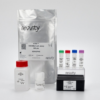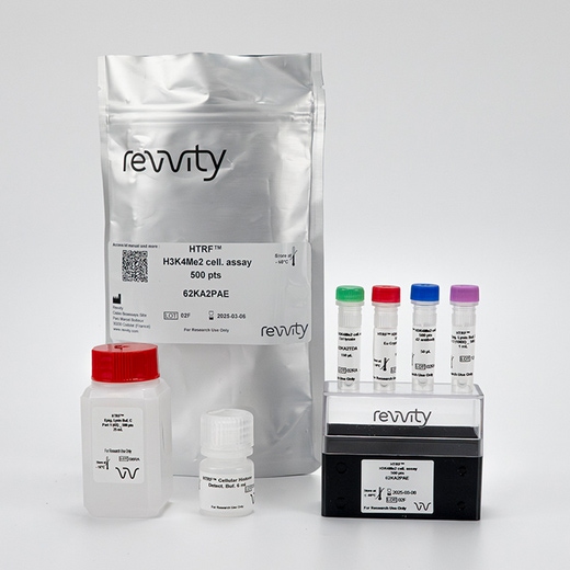

HTRF EPIgeneous 2-methyl K4 Histone H3 Detection Kit, 10,000 Assay Points


HTRF EPIgeneous 2-methyl K4 Histone H3 Detection Kit, 10,000 Assay Points


This cell-based assay format enables the detection of the H3K4Me2 motif, as a result of histone H3 modifications by SET or LSD1/2 enzymes among others.
| Feature | Specification |
|---|---|
| Application | Biochemical Enzymatic Assay |
| Sample Volume | 120 µL |
This cell-based assay format enables the detection of the H3K4Me2 motif, as a result of histone H3 modifications by SET or LSD1/2 enzymes among others.


HTRF EPIgeneous 2-methyl K4 Histone H3 Detection Kit, 10,000 Assay Points


HTRF EPIgeneous 2-methyl K4 Histone H3 Detection Kit, 10,000 Assay Points


Product information
Overview
This cellular assay has been developed with optimized reagents and protocols for the direct detection of endogenous levels of H3K4Me2.
This HTRF® format uses a very specific antibody pair combined with a highly efficient nucleus extraction which enables the assay to be used with a variety of cell backgrounds, and for the study of important methyltransferases and demethylases targets, such as JARID1 and MLL1.
The H3K27Me3 assay can be used for adherent or suspension cells, primary or secondary screening, and inhibitor studies.
Specifications
| Application |
Biochemical Enzymatic Assay
|
|---|---|
| Brand |
EPIgeneous
|
| Detection Modality |
HTRF
|
| Molecular Modification |
Methylation
|
| Product Group |
Kit
|
| Sample Volume |
120 µL
|
| Shipping Conditions |
Shipped in Dry Ice
|
| Target Class |
Epigenetics
|
| Technology |
TR-FRET
|
| Therapeutic Area |
Metabolism/Diabetes
Neuroscience
Oncology & Inflammation
|
| Unit Size |
10,000 Assay Points
|
Video gallery

HTRF EPIgeneous 2-methyl K4 Histone H3 Detection Kit, 10,000 Assay Points

HTRF EPIgeneous 2-methyl K4 Histone H3 Detection Kit, 10,000 Assay Points

How it works
Assay principle
The dimethylation of Lysine 4 on histone H3 is detected in a sandwich assay format using 2 different specific antibodies, one labeled with Eu-Cryptate (donor) and the second with d2 (acceptor). When the dyes are in close proximity, the excitation of the donor with a light source triggers a Fluorescence Resonance Energy Transfer (FRET), which in turn fluoresces at a specific wavelength (665 nm). One conjugate binds to Histone H3 and the other binds to the K4Me2 mark. The specific signal modulates positively in proportion to trimethylation on Lysine 4.

Two-plate assay protocol
Cells are plated (stimulated) and lysed in the same culture plate and then transferred to the assay plate for the detection of H3K4Me2 by HTRF reagents. This protocol enables the cells viability and confluence to be monitored.

One-plate assay protocol
Detection of H3K4Me2 with HTRF reagents is performed in a single plate used for plating, stimulation and detection. No washing steps are required. This protocol, HTS designed, allows miniaturization while maintaining HTRF quality.

Analytical performance
Detection of H3K4Me2 on several derivated cancer cell lines
HTRF detection of H3K4Me2 mark was done using a two-plate assay protocol. Cells were seeded at various densities in a 96-well plate and incubated for 24 h before lysis. Lysates were then transfered to a 384-sv plate for detection. To ensure operation in the linear range of the kit and to achieve an optimal S/B, cell densities have to be optimized.

Antibody specificity
HeLa cells were seeded at a density of 15,000 cells per well in a 96-well plate for a 24 h incubation. Serial dilutions of Histone H3 derived peptides (from 10 µM down to a minimum of 1 nM) with various epigenetic methyl marks were added to the detection well before the addition of HTRF detection conjugates. H3K4Me1 peptide competed with low affinity (IC50 = 1.08 µM), while the H3K4Me2 peptide competed with high affinity with the system leading to an IC50 value of 26.7 nM showing the specificity of the kit.

Measurement of the methylation of H3K4 induced by HDAC inhibitor
HeLa cells were seeded at 12,500cells per well in a 96-well plate, then treated overnight with TSA, NaB and Apicidine, 3 well known HDAC inhibitors that induce the di-methylation of H3K4 (H3K4Me2) and a decrease of H3K9Me2. After lysis, lysates were transfered to a 384-sv plate for detection using a two-plate assay protocol.

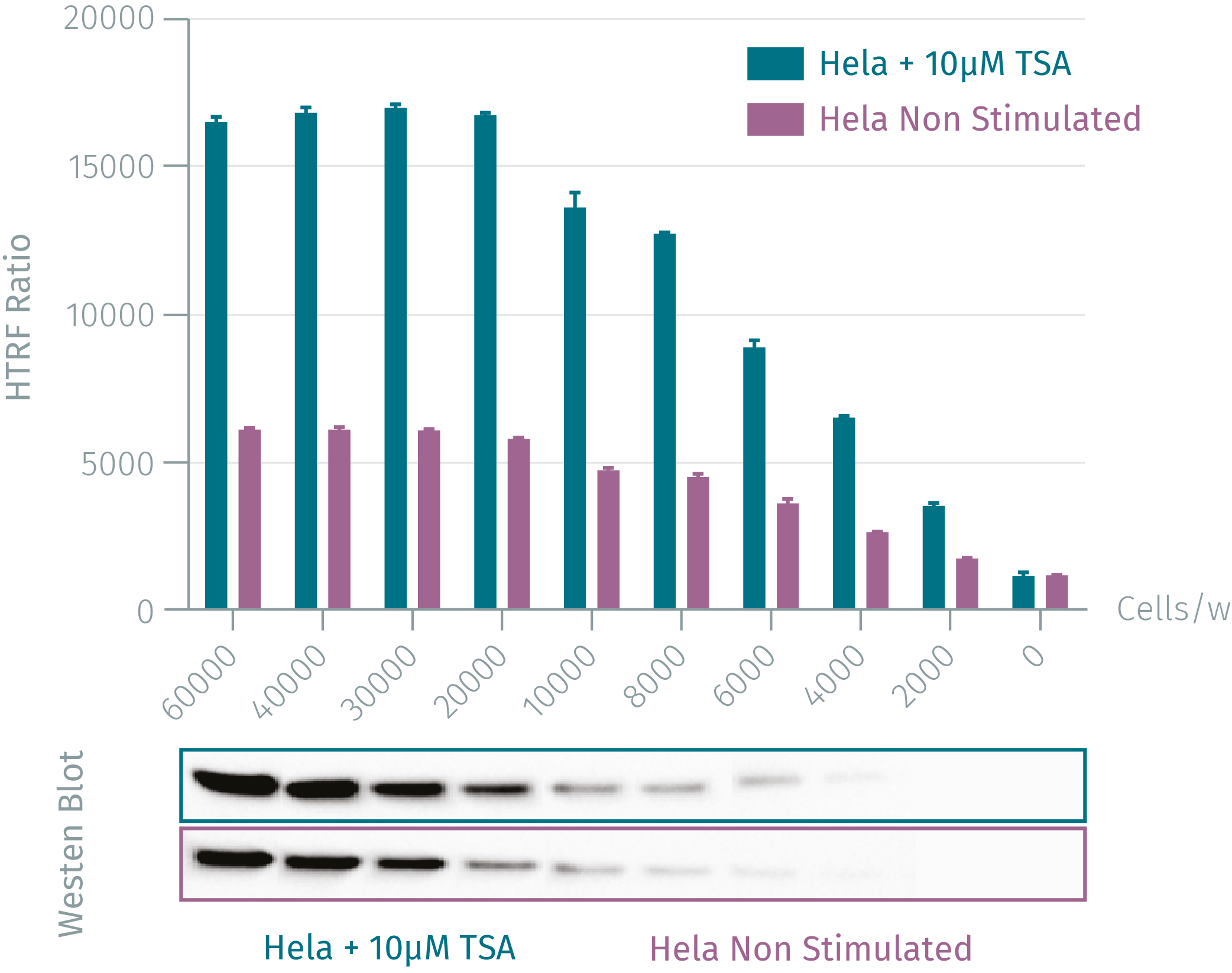
Western blot versus HTRF assay
Various concentrations of HeLa cells were grown in 96w plate at 37°C, 5% CO2 for 16 hours, with and without 10 µM TSA. After medium removal, the cells were lysed with Supplemented lysis buffer C. Each cell concentration was dispensed and analyzed side-by-side by Western Blot (sample vol = 13 µL) and by HTRF EPIgeneous H3K4Me2 assay (sample vol =10 µL). By using HTRF EPIgeneous H3K4Me2 assay only 2,000 cells are sufficient for minimal signal detection, while 6,000 cells are needed for a Western Blot signal.

Resources
Are you looking for resources, click on the resource type to explore further.
This guide provides you an overview of HTRF applications in several therapeutic areas.
Loading...


How can we help you?
We are here to answer your questions.


