


US
Revvity Sites Globally
Select your location.
*e-commerce not available for this region.

HTRF Human Total MerTK Detection Kit, 500 Assay Points

This HTRF kit allows for the cell-based quantitative detection of Total MerTK.
For research use only. Not for use in diagnostic procedures. All products to be used in accordance with applicable laws and regulations including without limitation, consumption & disposal requirements under European REACH regulations (EC 1907/2006).
Product Variants
Product information
Overview
MerTK (Myeloid-Epithelial-Reproductive Tyrosine Kinase) is a transmembrane tyrosine kinase receptor predominantly expressed in dendritic cells, macrophages, epithelial cells, and cells of the reproductive tract. When stimulated by ligands such as Protein S or Gas6, activated MerTK confers a survival advantage to cancer cells by promoting their survival, enhancing migration, and inhibiting apoptosis. The overexpression of MerTK is implicated in a wide range of cancers, making it an intriguing target for cancer therapies research.
Specifications
| Assay Target Class |
Phospho-protein
|
|---|---|
| Brand |
HTRF
|
| Buffer |
lysis buffer 4
|
| Detection Method |
HTRF
|
| Species |
Human
|
| Shipping Conditions |
Shipped in Dry Ice
|
| Therapeutic Area |
Inflammation
Oncology
|
| Unit Size |
500 Assay Points
|
How it works
Total MerTK assay principle
The Total MerTK assay quantifies the expression level of MerTK in a cell lysate. Unlike Western Blot, the assay is entirely plate-based and does not require gels, electrophoresis, or transfer. The Total MerTK assay uses two labeled antibodies, one coupled to a donor fluorophore and the other to an acceptor. Both antibodies are highly specific for a distinct epitope on the protein. In the presence of MerTK in a cell extract, the addition of these conjugates brings the donor fluorophore into close proximity with the acceptor and thereby generates a FRET signal. Its intensity is directly proportional to the concentration of the protein present in the sample and provides a means of assessing the protein's expression under a no-wash assay format.
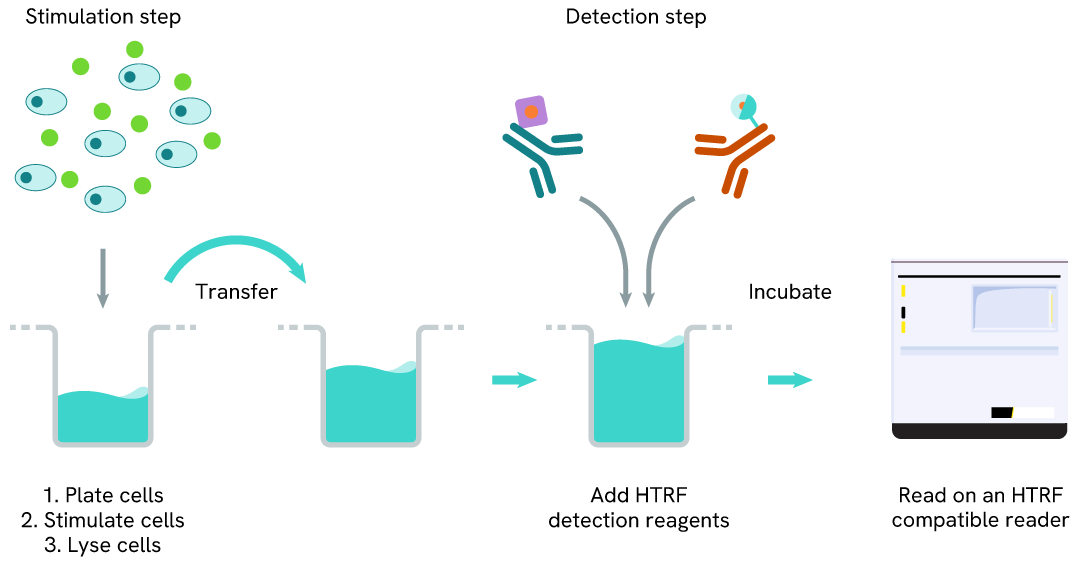
Total MerTK two-plate assay protocol
The two-plate protocol involves culturing cells in a 96-well plate before lysis, then transferring lysates into a 384-well low volume detection plate before the addition of HTRF Total MerTK detection reagents. This protocol allows for the cells' viability and confluence to be monitored.
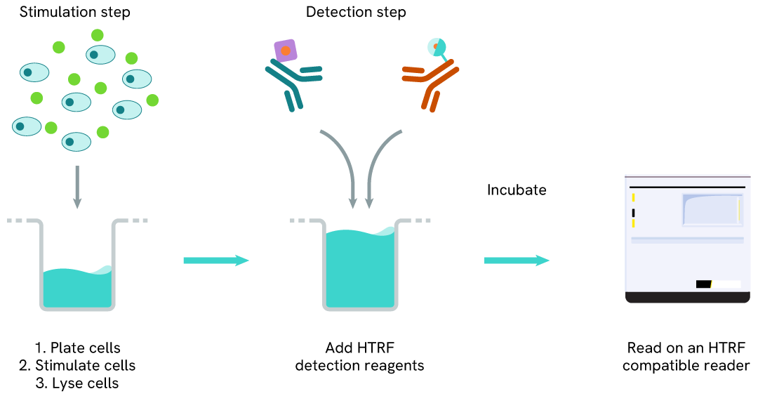
Total MerTK one-plate assay protocol
Detection of Total MerTK with HTRF reagents can be performed in a single plate used for culturing, stimulation, and lysis. No washing steps are required. This HTS designed protocol facilitates miniaturization while maintaining robust HTRF quality.
Assay validation
Induction of phospho-MerTK (Tyr749) in endogenous and overexpressed MerTK cellular models
HepG2 cells were seeded in a 96-well culture-treated plate (100,000 cells/well) in complete culture medium, and incubated overnight at 37°C, 5% CO2. The cells were treated for 30 minutes with 100 µM of Pervanadate.
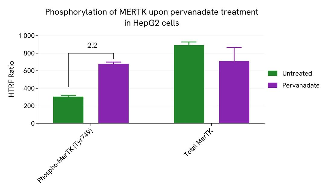
After treatment, the cells were lysed with 50 µL of supplemented lysis buffer #4 for 30 minutes at RT under gentle shaking. For the detection step, 16 µL of cell lysate were transferred into a 384-well low volume white microplate and 4 µL of the HTRF Phospho-MerTK (Tyr749) or Total MerTK detection reagents were added. The HTRF signal was recorded after an overnight incubation.
As expected, Pervanadate induced an increase in the level of Phospho-MerTK (Tyr749).
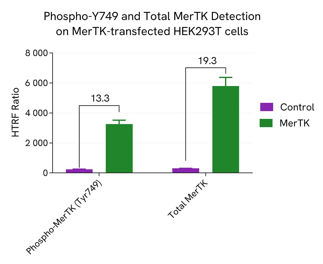
HEK293T cells were seeded in a 96-well culture-treated plate (12,500 cells/well) in complete culture medium, and incubated overnight at 37°C, 5% CO2. The cells were transfected with MerTK plasmid using DharmaFECT kb (Horizon Discovery) or with an empty plasmid as control.
After 24h of incubation, the cells were lysed with 50 µL of supplemented lysis buffer #4 for 30 minutes at RT under gentle shaking. For the detection step, 16 µL of cell lysate were transferred into a 384-well low volume white microplate and 4 µL of the HTRF Phospho-MerTK (Tyr749) or Total MerTK detection reagents were added. The HTRF signal was recorded after an overnight incubation. As anticipated, the increased levels of MerTK without treatment led to its autophosphorylation, allowing for the detection of Phospho and Total MerTK.
Inhibition of phospho-MerTK (Tyr749) in endogenous and overexpressed MerTK cellular models
HepG2 cells were seeded in a 96-well culture-treated plate (100,000 cells/well) in complete culture medium, and incubated overnight at 37°C, 5% CO2. The cells were treated for 1 hour with increasing concentrations of UNC1062 (a MerTK inhibitor), and 5 µM of Pervanadate were added 30 minutes before the end of the treatment. The cells were lysed with 50 µL of supplemented lysis buffer #4 for 30 minutes at RT under gentle shaking.
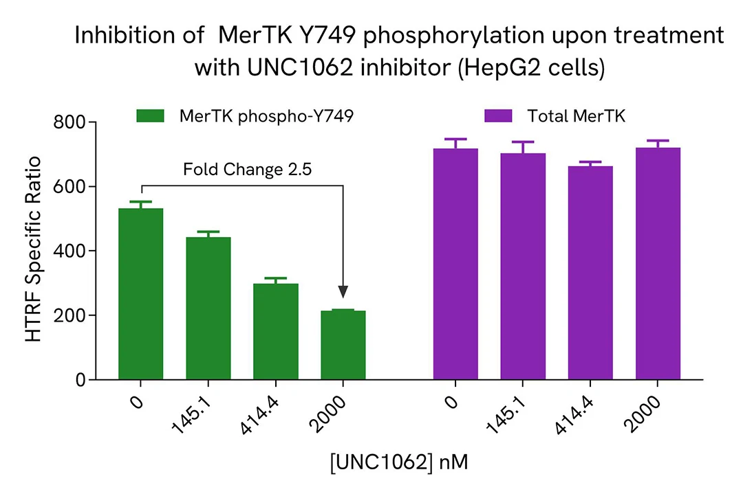
For the detection step, 16 µL of cell lysate were transferred into a 384-well low volume white microplate and 4 µL of the HTRF Phospho-MerT (Tyr749) or Total MerTK detection reagents were added. The HTRF signal was recorded after an overnight incubation.
As anticipated, the MerTK inhibitor UNC1062 led to a dose-dependent reduction in MerTK phosphorylation, while not affecting the expression level of the receptor.
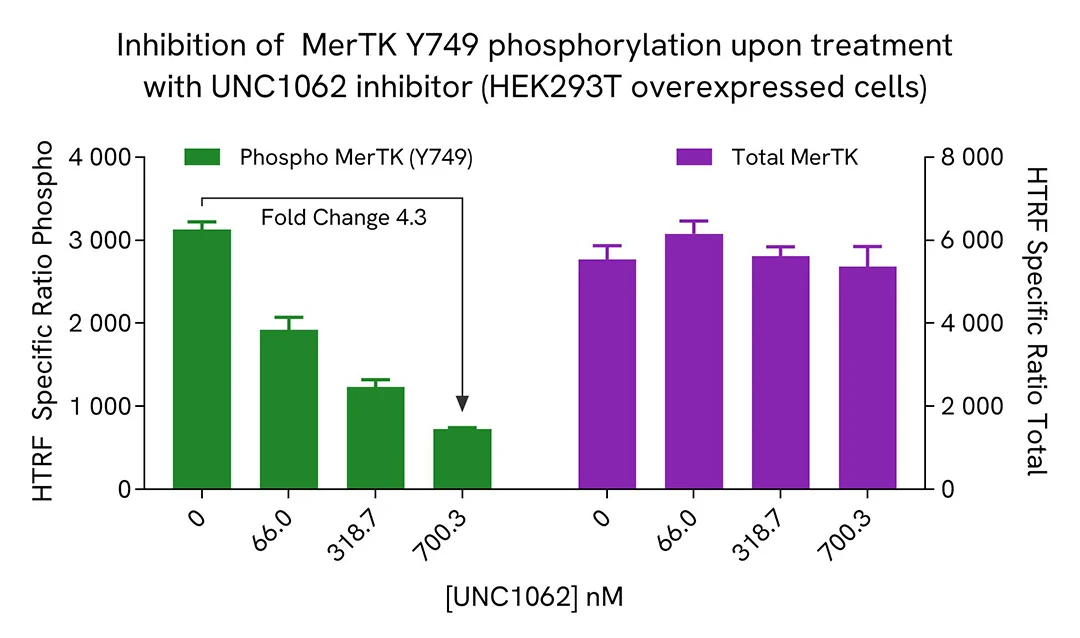
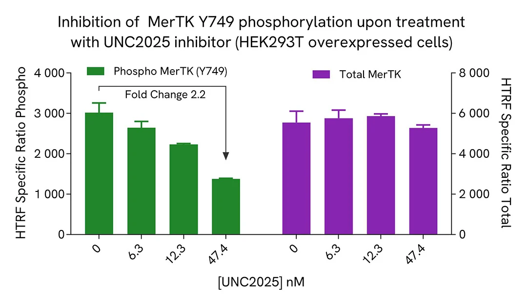
HEK293T cells were seeded in a 96-well culture-treated plate (12,500 cells/well) in complete culture medium, and incubated overnight at 37°C, 5% CO2. The cells were transfected with MerTK plasmid using DharmaFECT kb (Horizon Discovery). After 24h of incubation, the cells were treated for 1 hour with increasing concentrations of UNC1062 or UNC2025 (MerTK inhibitors).
The cells were lysed with 50 µL of supplemented lysis buffer #4 for 30 minutes at RT under gentle shaking. For the detection step, 16 µL of cell lysate were transferred into a 384-well low volume white microplate and 4 µL of the HTRF Phospho-MerTK (Tyr749) or Total MerTK detection reagents were added. The HTRF signal was recorded after an overnight incubation.
As expected, both MerTK inhibitors led to a dose-dependent reduction in MerTK phosphorylation, while not affecting the expression level of the receptor.
Selectivity of Total-MerTK assay using transfection of members of the TAM family
HEK293T cells were grown in a T175 flask in complete culture medium at 37°C - 5% CO2 until 80% confluence. Transfection of MerTK, Axl, or Tyro3, as well as a negative control, was carried out using DharmaFECT kb (Revvity’s Horizon Discovery products). After 24h of incubation, the cells were treated for 30 min with 100 µM of Pervanadate before being lysed with 3 mL of supplemented lysis buffer #4 (1X) for 30 minutes at RT under gentle shaking.
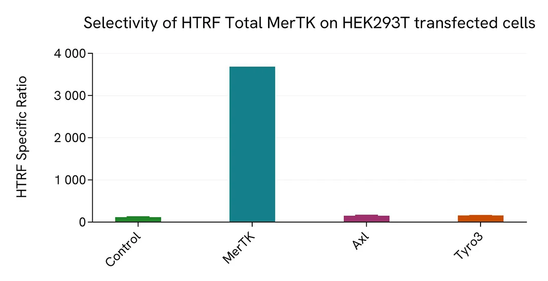
After cell lysis, 16 µL of lysates, diluted 1/16 in supplemented lysis buffer #4 (1X) to be in the linear range of the assay, were transferred into a 384-well low volume white microplate and 4 µL of the HTRF Total-MerTK detection reagents were added. The HTRF signal was recorded after an overnight incubation.
Cell transfection with MerTK alone resulted in the detection of Total-MerTK compared to the negative control. In contrast, transfection of Axl or Tyro3 did not lead to any signal increase, demonstrating that the Total-MerTK assay is specific for MerTK and, as expected, does not cross-react with other members of the TAM family.
Assessment of Total and phospho-MerTK (Tyr749) level in various cell lines
HepG2 cells with endogeneous expression of MerTK and Hek293T cells overexpressing MerTK were seeded at respectively 100,000 and 12,500 cells/well in a 96-well microplate. After 24h of incubation, the cells were treated for 30 min with 100 µM of Pervanadate before being lysed with 50 µL of supplemented lysis buffer #4 for 30 minutes at RT under gentle shaking.
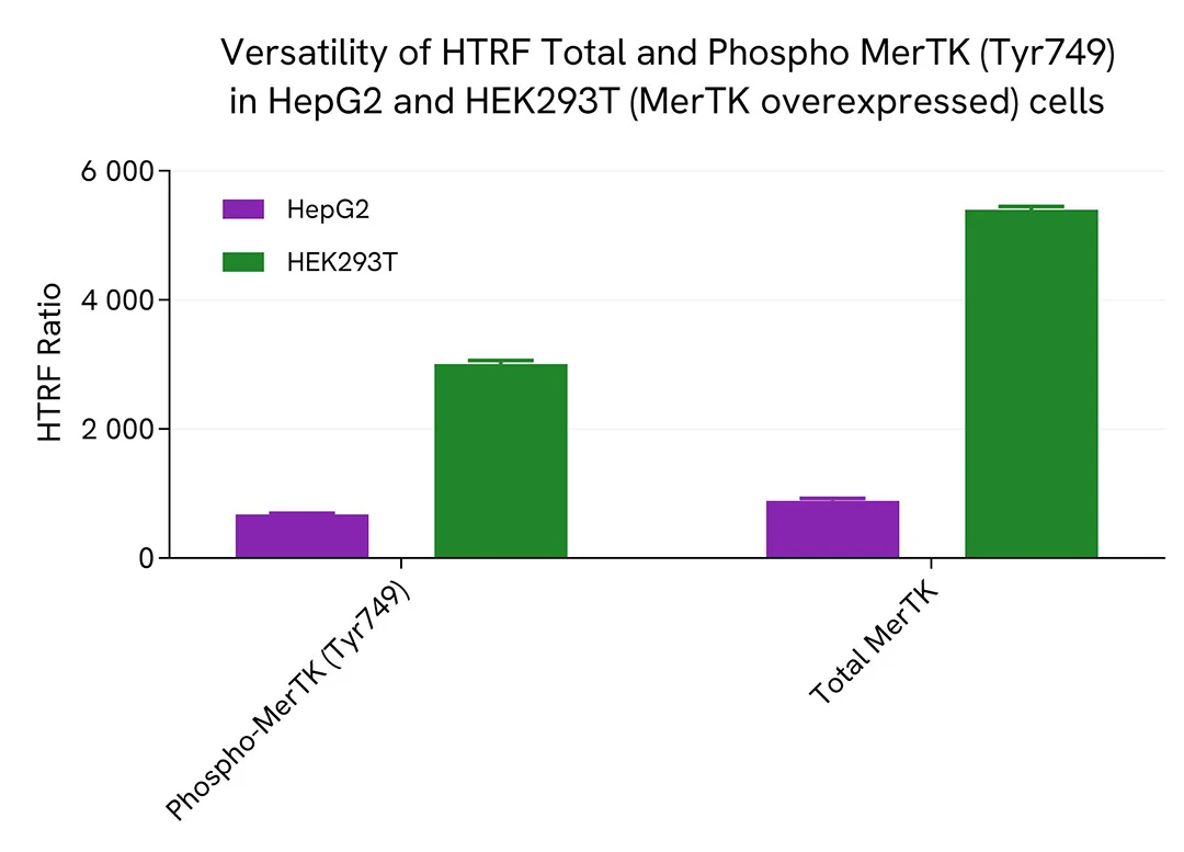
16 µL of lysate were transferred into a 384-well low volume white microplate before the addition of 4 µL of the HTRF Phospho-MerTK (Tyr749) or HTRF Total MerTK detection reagents. The HTRF signal was recorded after an overnight incubation.
The HTRF Total MerTK assay efficiently detected Total MerTK in various cellular models expressing dvarying levels of the protein.
HTRF Total MerTK assay compared to Western Blot
HEK293 cells were grown in a T175 flask in complete culture medium at 37°C - 5% CO2 until 80% confluence. Transfection of MerTK plasmid was performed with DharmaFECT kb (Revvity’s Horizon Discovery products). After 24h of incubation, the cells were lysed with 3 mL of supplemented lysis buffer #4 (1X) for 30 minutes at RT under gentle shaking.
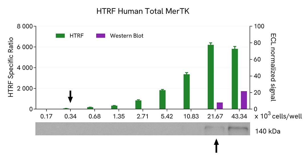
Serial dilutions of the cell lysate were performed using supplemented lysis buffer, and 16 µL of each dilution were transferred into a low volume white microplate before the addition of 4 µL of HTRF Total-MerTK detection reagents. Equal amounts of lysates were used for a side-by-side comparison between HTRF and Western Blot.
Using the HTRF Total MerTK assay, 340 cells/well were enough to detect a significant signal, while 21670 cells were needed to obtain a minimal chemiluminescent signal using Western Blot. Therefore, in these conditions, the HTRF Total MerTK assay was 64 times more sensitive than the Western Blot technique.
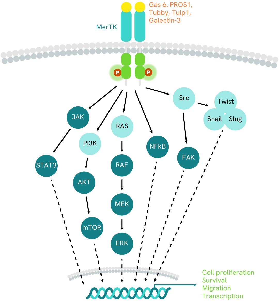
Simplified pathway
MerTK signaling pathway
MerTK, also known as Mer receptor tyrosine kinase, belongs to the TAM family of tyrosine kinase receptors. It plays crucial roles in various cellular mechanisms, including inflammatory responses, cell survival, phagocytosis, and the clearance of apoptotic cell debris. Activation of MerTK occurs when it interacts with specific ligands such as Gas6, protein S, and Galectins. These ligands act as bridges between the receptor and cellular debris generated by nearby apoptotic cells. Upon activation, MerTK undergoes dimerization and autophosphorylation at specific intracellular tyrosine residues. These phosphorylated sites serve as docking points for recruiting and activating downstream signaling proteins. The downstream pathways influenced by MerTK include the PI3K/AKT pathway (involving the mTOR transcription factor), the MAPK pathway (contributing to cell proliferation and differentiation), and the JAK/STAT pathway (relying on STAT transcription factors to induce inflammatory responses). In summary, MerTK orchestrates essential cellular processes by integrating signals from its ligands and initiating downstream pathways.
Resources
Brochure
HTRF assays and reagents catalog
Discover the versatility and precision of Homogeneous Time-Resolved Fluorescence (HTRF) technology. Our HTRF portfolio offers a...

SDS, COAs, Manuals and more
Interested in technical resources related to this product? We've curated them in dedicated sections for your convenience. Click the links below to explore further.


How can we help you?
We are here to answer your questions.

