

HTRF Total Myd88 Detection Kit, 500 Assay Points
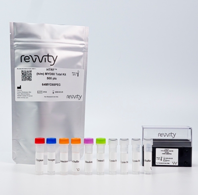
HTRF Total Myd88 Detection Kit, 500 Assay Points
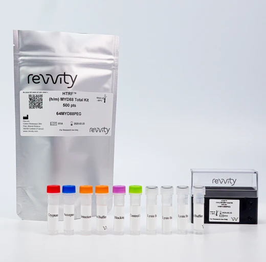


This HTRF kit enables the cell-based quantitative detection of Total Myd88.
For research use only. Not for use in diagnostic procedures. All products to be used in accordance with applicable laws and regulations including without limitation, consumption and disposal requirements under European REACH regulations (EC 1907/2006).
| Feature | Specification |
|---|---|
| Application | Cell Signaling |
| Sample Volume | 16 µL |
This HTRF kit enables the cell-based quantitative detection of Total Myd88.
For research use only. Not for use in diagnostic procedures. All products to be used in accordance with applicable laws and regulations including without limitation, consumption and disposal requirements under European REACH regulations (EC 1907/2006).


HTRF Total Myd88 Detection Kit, 500 Assay Points


HTRF Total Myd88 Detection Kit, 500 Assay Points


Product information
Overview
Myeloid differentiation factor 88 (MyD88) is a central adaptor for Toll-like or interleukin-1 Receptors (TLR/IL-1R) signaling that mediates initiation of the innate immune response and production of the proinflammatory cytokines that restrain pathogens and activate adaptive immunity. Although MyD88 is crucial for a host to prevent pathogenic infection, misregulation of its abundance might lead to autoimmune diseases. Degradation of MyD88 is a key canonical mechanism for terminating cytokine production.
Specifications
| Application |
Cell Signaling
|
|---|---|
| Brand |
HTRF
|
| Detection Modality |
HTRF
|
| Lysis Buffer Compatibility |
Lysis Buffer 1
Lysis Buffer 2
Lysis Buffer 3
Lysis Buffer 4
|
| Molecular Modification |
Total
|
| Product Group |
Kit
|
| Sample Volume |
16 µL
|
| Shipping Conditions |
Shipped in Dry Ice
|
| Target Class |
Phosphoproteins
|
| Technology |
TR-FRET
|
| Unit Size |
500 Assay Points
|
How it works
Total Myd88 assay principle
The HTRF Total Myd88 assay quantifies the expression level of Myd88 in a cell lysate. Unlike Western Blot, the assay is entirely plate-based, and does not require gels, electrophoresis, or transfer. The Total Myd88 assay uses two labeled antibodies, one coupled to a donor fluorophore, the other to an acceptor. Both antibodies are highly specific for a distinct epitope on the protein. In presence of Myd88 in a cell extract, the addition of these conjugates brings the donor fluorophore into close proximity with the acceptor, and thereby generates a FRET signal. Its intensity is directly proportional to the concentration of the protein present in the sample, and provides a means of assessing the protein’s expression under a no-wash assay format.

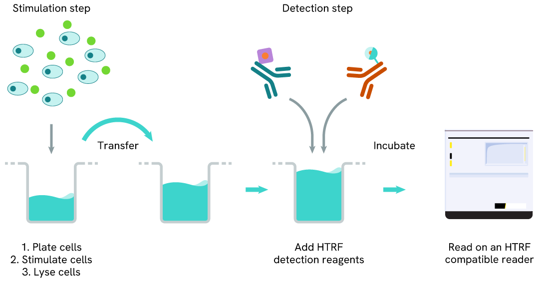
Total Myd88 two-plate assay protocol
The 2-plate protocol involves culturing cells in a 96-well plate before lysis, then transferring lysates into a 384-well low volume detection plate before the addition of the Total Myd88 HTRF detection reagents. This protocol enables the cells' viability and confluence to be monitored.
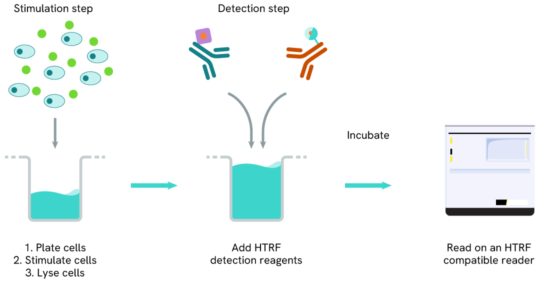
Human Total Myd88 one-plate assay protocol
Detection of Total Myd88 with HTRF reagents can be performed in a single plate used for culturing, stimulation, and lysis. No washing steps are required. This HTS-designed protocol enables miniaturization while maintaining robust HTRF quality.
Assay validation
HTRF Total MYD88 expression on THP-1 derived marcophages using LPS
THP1 cells were plated at 50, 000 cells/well and treated with 100ng/mL PMA for 24h, followed by 1µg/mL LPS for 24h or 1h to induce modulation of MyD88, IL-1B, TNF alpha, or phospho-NFkB, respectively.
Cells treated for 1h or 24h with LPS were then lysed with 50 µl of supplemented Lysis Buffer#1 (1X) for 30 min at RT under gentle shaking.
16 µL of lysate (24h treatment condition) were transferred into a 384-well low volume white microplate and 4 µL of the HTRF Total MyD88 detection antibodies were added. An additional 4 µL of lysate (supplemented with 12 µL diluent #8) were also transferred into the microplate to check the total protein content with the alpha-tubulin housekeeping Cellular Kit (64ATUBPET/G/H). 16µL of the respective supernatants were also transferred into the microplate to be analyzed with the Human TNF alpha (62HTNFAPET/G/H) and the Human IL-1beta (62HIL1B2PET/G/H) assays.
Additionally, 16 µL of lysate (1h treatment condition) were analyzed with phospho-NFkB (64NFBPET/G/H) and Total NFkB (64NFTPEG/H) assays. HTRF signals were recorded after an overnight incubation at room temperature.
Our results show an upregulation of MyD88 expression level induced by LPS exposure which was associated with a decrease in the Alpha Tubulin level, likely due to a cytotoxicity effect (data not shown). In addition, LPS induced NFkB phosphorylation and the subsequent release of the proinflammatory cytokines IL-1B and TNF alpha.


Specificity of HTRF Total MYD88 assay
The total MyD88 protein levels were assessed with the HTRF Human Total MyD88 kit in HAP1 cells (WT) and different HAP1 cell lines Knocked-Out for MyD88. Cell density was optimized beforehand to ensure HTRF detection within the dynamic range of the kit (data not shown).
The different cell lines were cultured in a 96-well plate (50,000 cells/well) for 24 hours at 37°C, 5% CO2. The cells were then lysed with 50 µL of supplemented lysis buffer #1 (1X) for 30 minutes at RT under gentle shaking, and 16 µL of cell lysate were transferred into a low volume white microplate followed by 4 µL of premixed detection reagents. The HTRF signal was recorded after an overnight incubation at RT.
In HAP1 KO MyD88 cells, the HTRF signal was equivalent to the non-specific signal (dotted line), indicating a complete Myd88 gene silencing, whereas the Myd88 level was well detected in the other cell line, as expected.
Catalog cell line references (Horizon Discovery): HAP1 Wt #C631; HAP1 KO MyD88 # HZGHC002929c003 and #HZGHC002929c011

Validation on various Human and Mouse cell lines
The suspension cell lines THP-1 (acute monocytic leukemia), Ramos (B lymphocyte), and KG-1 (acute myeloid leukemia) were dispensed into a 96-well plate and lysed with 10 µL of supplemented lysis buffer #1 (4X) for 30 min at RT under gentle shaking (performed following the suspension cell protocol).
The adherent cell lines PC-3 (prostatic adenocarcinoma) and NIH-3T3 (murine embryonic fibroblast) were plated in a 96-well culture plate and incubated for 24 hours at 37°C, 5% CO2. After medium removal, cells were lysed with 50 µL of supplemented lysis buffer #1 (1X) for 30 min at RT under gentle shaking.
The MyD88 expression level was assessed with the HTRF Total MyD88 kit. Briefly, 16 µL of cell lysate were transferred into a low volume white microplate, followed by 4 µL of premixed HTRF detection reagents. The HTRF signal was recorded after an overnight incubation at RT. The dotted line corresponds to the non-specific HTRF signal. Note that the cell density was optimized beforehand to ensure HTRF detection within the dynamic range of the kit (data are shown for 200,000 cells/well)
The HTRF Total MyD88 assay efficiently detected endogenous MyD88 in various cellular models expressing different levels of the protein.

Comparison between HTRF and Western Blot sensitivity for Total Myd88
Ramos cells were cultured in a T175 flask in complete medium at 37°C, 5% CO2. After medium removal, the cells were lysed with 3 mL of supplemented lysis buffer #1 (1X) for 30 min at RT under gentle shaking.
Serial dilutions of the cell lysate were performed using supplemented lysis buffer #1 (1X), and 16µL of each dilution were transferred into a 384-well small volume microplate before the addition of 4µL of HTRF Total MyD88 detection antibodies. HTRF signals were recorded after an overnight incubation.
Equal amounts of lysates were loaded into a gel for a side-by-side comparison between HTRF and Western Blot.
In these conditions, the HTRF Total MyD88 assay is 4-fold more sensitive than the Western Blot technique.

Simplified pathway
MyD88, a downstream adaptor protein of TLR, is critical in the signal transduction of the TLR signaling pathway. Upon binding of PAMPs or DAMPs to TLRs , the death domain (DD) of MyD88 interacts with the DD of IL-1 receptor-associated kinase-4 (IRAK-4) and forms the MyD88-IRAK-4 complex, which recruits IRAK-1 and IRAK-2, resulting in the phosphorylation of IRAKs. After phosphorylation, IRAKs interact with TRAF6 leading to the activation of the NFκB and MAPK pathways and resulting in the transcription of pro-inflammatory cytokines.

Resources
Are you looking for resources, click on the resource type to explore further.
Discover the versatility and precision of Homogeneous Time-Resolved Fluorescence (HTRF) technology. Our HTRF portfolio offers a...
This document includes detailed tables listing HTRF™, AlphaLISA™ SureFire® Ultra™, and Alpha SureFire® Ultra™ Multiplex assays...


How can we help you?
We are here to answer your questions.


