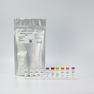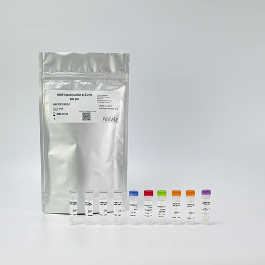

HTRF Human & Mouse Phospho-Cofilin (Ser3) Detection Kit, 500 Assay Points


HTRF Human & Mouse Phospho-Cofilin (Ser3) Detection Kit, 500 Assay Points


This HTRF kit enables the cell-based quantitative detection of Cofilin phosphorylation at Ser3.
For research use only. Not for use in diagnostic procedures. All products to be used in accordance with applicable laws and regulations including without limitation, consumption and disposal requirements under European REACH regulations (EC 1907/2006).
| Feature | Specification |
|---|---|
| Application | Cell Signaling |
| Sample Volume | 16 µL |
This HTRF kit enables the cell-based quantitative detection of Cofilin phosphorylation at Ser3.
For research use only. Not for use in diagnostic procedures. All products to be used in accordance with applicable laws and regulations including without limitation, consumption and disposal requirements under European REACH regulations (EC 1907/2006).


HTRF Human & Mouse Phospho-Cofilin (Ser3) Detection Kit, 500 Assay Points


HTRF Human & Mouse Phospho-Cofilin (Ser3) Detection Kit, 500 Assay Points


Product information
Overview
The kit is designed for the rapid detection of Phospho Cofilin Ser3 in cell supernatant and whole cells. Actin-binding proteins are abundant cellular proteins that regulate cell function by mediating actin polymerization and remodeling. Cofilin, known as a regulator of actin filament dynamics, is a small ~21kDA protein that is ubiquitously expressed in all vertebrates and freely diffuses in eukaryotic cells. Phosphorylation at Ser-3 by kinases attenuates cofilinâ's actin- binding activity. In contrast, dephosphorylation at Ser-3 enhances cofilin-induced actin depolymerization.
Specifications
| Application |
Cell Signaling
|
|---|---|
| Brand |
HTRF
|
| Detection Modality |
HTRF
|
| Lysis Buffer Compatibility |
Lysis Buffer 4
Lysis Buffer 5
|
| Molecular Modification |
Phosphorylation
|
| Product Group |
Kit
|
| Sample Volume |
16 µL
|
| Shipping Conditions |
Shipped in Dry Ice
|
| Target Class |
Phosphoproteins
|
| Target Species |
Human
Mouse
|
| Technology |
TR-FRET
|
| Unit Size |
500 Assay Points
|
Video gallery

HTRF Human & Mouse Phospho-Cofilin (Ser3) Detection Kit, 500 Assay Points

HTRF Human & Mouse Phospho-Cofilin (Ser3) Detection Kit, 500 Assay Points

How it works
Phospho-Cofilin (Ser3) assay principle
The Phospho-Cofilin (Ser3) assay measures Cofilin when phosphorylated at Ser3. Unlike Western Blot, the assay is entirely plate-based and does not require gels, electrophoresis, or transfer. The assay uses 2 antibodies, one labeled with a donor fluorophore and the other with an acceptor. The first antibody was selected for its specific binding to the phosphorylated motif on the protein, the second for its ability to recognize the protein independently of its phosphorylation state. Protein phosphorylation enables an immune-complex formation involving both labeled antibodies and which brings the donor fluorophore into close proximity to the acceptor, thereby generating a FRET signal. Its intensity is directly proportional to the concentration of phosphorylated protein present in the sample, and provides a means of assessing the protein’s phosphorylation state under a no-wash assay format.

Phospho-Cofilin (Ser3) two-plate assay protocol
The 2-plate protocol involves culturing cells in a 96-well plate before lysis, then transferring lysates to a 384-well low volume detection plate before the addition of Phospho-Cofilin (Ser3) HTRF detection reagents. This protocol enables the cells' viability and confluence to be monitored.

Phospho-Cofilin (Ser3) one-plate assay protocol
Detection of Phosphorylated Cofilin (Ser3) with HTRF reagents can be performed in a single plate used for culturing, stimulation, and lysis. No washing steps are required. This HTS designed protocol enables miniaturization while maintaining robust HTRF quality.

Assay validation
HTRF Phospho-Cofilin (Ser3) / Total Cofilin modulation using LIMK1 inhibitor and Staurosporine
HeLa cells were plated at 10,000 cell/well in complete culture medium, and incubated 24 hours at 37°C, 5% CO2. Cells were treated with increasing concentrations of Staurosporine or LIMK1 kinase (TH257) for respectively 1 or 2 hours at 37°C, 5% CO2. Supernatant was then discarded and 50µl of supplemented lysis buffer#4 was added to each well. The lysis step was done under gentle shaking for 30 minutes. Lysates were transferred into a 384 well plate under 16µl per well, and 4µL of Total or Phospho Cofilin Ser3 detection reagents were dispensed. The plate was read after an overnight incubation.
As expected, both compounds induced a decrease in phosphorylated Cofilin at Ser3 upon treatment, while the Total Cofilin expression level was not modulated.


HTRF Phospho-Cofilin (Ser3) / Total Cofilin on neuronal cells
Human neuronal cells were plated at 10,000 cell/well in complete culture medium, and incubated 24 hours at 37°C, 5% CO2. Cells were treated with increasing concentrations of Staurosporine or LIMK1 kinase (TH257) for respectively 1 or 2 hours at 37°C, 5% CO2. Supernatant was then discarded, and 50µl of complemented lysis buffer#4 were added to each well. The lysis step was done under gentle shaking for 30 minutes. Lysates were transferred into a 384 well plate under 16µl per well, and 4µL of total or Phospho Cofilin Ser3 detection reagents were dispensed. The plate was read after an overnight incubation.
As expected, both compounds induced a decrease in phosphorylated Cofilin at Ser3 upon treatment, while the Total Cofilin expression level was not modulated.


HTRF Specificity of Cofilin Phospho-S3 assay using SiRNA
Hela cells were plated at 5,000 cell/well in complete culture medium, and incubated 24 hours at 37°C, 5% CO2. Cells were transfected with 50nmol of siRNA against Cofilin 1 and/or Cofilin 2 for 24 hours at 37°C, 5% CO2. Transfection medium was then removed, and cell culture medium was added on top of the cells for the next 24 hours at 37°C, 5% CO2. Supernatant was then discarded and 50µl of complemented lysis buffer#4 were added to each well. The lysis step was done under gentle shaking for 30 minutes. Lysates were transferred into a 384 well plate under 16µl per well and 4µL of Phospho Cofilin Ser3 detection reagents were dispensed. The plate was read after an overnight incubation.
Cofilin 1 siRNA induced a 90% signal decrease in the phospho Cofilin 1 detection, while this was not the case for Cofilin 2. The data demonstrate that HTRF Phospho Cofilin 1 is specific for the detection of the Cofilin 1 protein, and does not cross-react with Cofilin 2.

Cofilin Phospho-S3 assay versatility on human & mouse cell lines
Human HeLa and HepG2 cells and mouse NIH-3T3 cells were plated at 12,500 cells/well in complete culture medium, and incubated 24 hours at 37°C, 5% CO2. Supernatant was then discarded, and 50µl of supplemented lysis buffer#4 were added to each well. The lysis step was done under gentle shaking for 30minutes. Lysates were transferred into a 384 well plate under 16µl per well and 4µL of Phospho Cofilin Ser3 detection reagents were dispensed. The plate was read after an overnight incubation.
Phospho Cofilin protein was efficiently detected in various human and mouse cellular models.

Comparison between HTRF and WB sensitivity for Cofilin phospho-S3
HeLa cells were cultured in a T175 flask in complete culture medium at 37°C, 5% CO2. After a 48h incubation, cells were lysed with 3mL of supplemented lysis buffer #4 (1X), for 30 minutes at RT under gentle shaking.
After a first dilution by 10, serial dilutions of the cell lysate were performed using supplemented lysis buffer, and 16 µL of each dilution were transferred into a low volume white microplate before the addition of 4 µL of HTRF Phospho-Cofilin Ser3 detection reagents. Equal amounts of lysates were used for a side-by-side comparison between HTRF and Western Blot.
In these conditions, the HTRF Phospho-Cofilin Ser3 assay is 32 times more sensitive than the Western Blot technique.

Simplified pathway
Cofilin Signaling Pathway
Actin-binding proteins are abundant cellular proteins that regulate cell function by mediating actin polymerization and remodeling. Cofilin, known as a regulator of actin filament dynamics, is a small ~21kDA protein that is ubiquitously expressed in all vertebrates and freely diffuses in eukaryotic cells. Cofilin promotes the conversion of actin filaments by enhancing F- actin depolymerization and inhibiting G-actin polymerization, which are essential in the actin filament dynamics of eukaryotes. Phosphorylation at Ser-3 by kinases attenuates cofilin’s actin- binding activity. In contrast, dephosphorylation at Ser-3 enhances cofilin-induced actin depolymerization. Cofilin functions are also modulated by various binding partners or reactive oxygen species.
Although the mechanism of cofilin-mediated actin dynamics has been known for decades, recent research works have unveiled the profound impacts of cofilin dysregulation in neurodegenerative pathophysiology. For instance, an oxidative stress-induced increase in cofilin dephosphorylation has been linked to the accumulation of tau tangles and amyloid-beta plaques in Alzheimer’s Disease. In Parkinson’s Disease, cofilin activation by silencing its upstream kinases increases α-synuclein-fibril entry into the cell. Cofilin is also over expressed in cancer cells, promoting cell motility and acting as an important regulator of cancer metastasis.

Resources
Are you looking for resources, click on the resource type to explore further.
Discover the versatility and precision of Homogeneous Time-Resolved Fluorescence (HTRF) technology. Our HTRF portfolio offers a...


How can we help you?
We are here to answer your questions.


