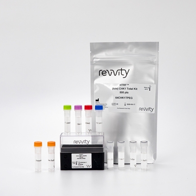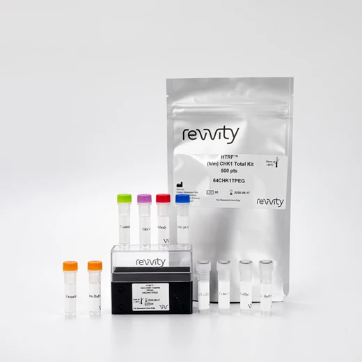

HTRF Human & Mouse Total CHK-1 Detection Kit, 10,000 Assay Points


HTRF Human & Mouse Total CHK-1 Detection Kit, 10,000 Assay Points


The Total CHK1 kit is designed to monitor the expression level of cellular CHK1, a Ser kinase involved in DNA damage response. This kit can be used as a normalization assay with our Phospho-CHK1 Ser 345 kit to enable optimal investigation of the ATR/CHK1 pathway.
For research use only. Not for use in diagnostic procedures. All products to be used in accordance with applicable laws and regulations including without limitation, consumption and disposal requirements under European REACH regulations (EC 1907/2006).
| Feature | Specification |
|---|---|
| Application | Cell Signaling |
| Sample Volume | 16 µL |
The Total CHK1 kit is designed to monitor the expression level of cellular CHK1, a Ser kinase involved in DNA damage response. This kit can be used as a normalization assay with our Phospho-CHK1 Ser 345 kit to enable optimal investigation of the ATR/CHK1 pathway.
For research use only. Not for use in diagnostic procedures. All products to be used in accordance with applicable laws and regulations including without limitation, consumption and disposal requirements under European REACH regulations (EC 1907/2006).


HTRF Human & Mouse Total CHK-1 Detection Kit, 10,000 Assay Points


HTRF Human & Mouse Total CHK-1 Detection Kit, 10,000 Assay Points


Product information
Overview
The Total-CHK1 cellular assay kit is designed to monitor the expression level of CHK1, whether phosphorylated or unphosphorylated.
It is compatible with our Phospho-CHK1 ser 345 kit, and enables the analysis of phosphorylated and total proteins from a single sample for a better readout of the ATR/CHK1 pathway.
Specifications
| Application |
Cell Signaling
|
|---|---|
| Automation Compatible |
Yes
|
| Brand |
HTRF
|
| Detection Modality |
HTRF
|
| Lysis Buffer Compatibility |
Lysis Buffer 1
Lysis Buffer 2
Lysis Buffer 4
|
| Molecular Modification |
Total
|
| Product Group |
Kit
|
| Sample Volume |
16 µL
|
| Shipping Conditions |
Shipped in Dry Ice
|
| Target Class |
Phosphoproteins
|
| Target Species |
Human
Mouse
|
| Technology |
TR-FRET
|
| Therapeutic Area |
Oncology & Inflammation
|
| Unit Size |
10,000 Assay Points
|
Video gallery

HTRF Human & Mouse Total CHK-1 Detection Kit, 10,000 Assay Points

HTRF Human & Mouse Total CHK-1 Detection Kit, 10,000 Assay Points

How it works
HTRF Total CHK1 assay principle
The Total-CHK1 assay quantifies the expression level of CHK1 in a cell lysate. Unlike Western Blot, the assay is entirely plate-based and does not require gels, electrophoresis, or transfer.
The Total-CHK1 assay uses two labeled antibodies: one coupled to a donor fluorophore, the other to an acceptor. Both antibodies are highly specific for a distinct epitope on the protein. In presence of CHK1 in a cell extract, the addition of these conjugates brings the donor fluorophore into close proximity with the acceptor and thereby generates a FRET signal. Its intensity is directly proportional to the concentration of the protein present in the sample, and provides a means of assessing the protein’s expression under a no-wash assay format.

HTRF Total-CHK1 2-plate assay protocol
The two-plate protocol involves culturing cells in a 96-well plate before lysis, then transferring lysates to a 384-well low volume detection plate before the addition of the Total-CHK1 HTRF detection reagents.
This protocol enables the cells' viability and confluence to be monitored.

Total-CHK1 1-plate assay protocol
Detection of Total CHK1 with HTRF reagents can be performed in a single plate used for culturing, stimulation, and lysis. No washing steps are required.
This HTS-designed protocol enables miniaturization while maintaining robust HTRF quality.

Assay validation
Human/Mouse CHK1 assay compatibility: Neocarzinostatin dose response curves
Human HEK293 cells or Mouse NIH/3T3 cells were plated in a 96-well culture-treated plate (100,000 cells/well) in complete culture medium, and incubated overnight at 37°C, 5% CO2. The cells were treated with a dose-response of Neocarzinostatin 2h at 37 °C, 5% CO2. After medium removal, the cells were lysed with 50 µl of supplemented lysis buffer #1 (1X) for 30 min at RT under gentle shaking. After cell lysis, 16 µL of lysate were transferred into a 384-well low volume white microplate and 4 µL of the HTRF Phospho CHK1 (Ser 345) or Total-CHK1 detection reagents were added. The HTRF signal was recorded after an overnight incubation at room temperature.
As expected, Neocarzinostatin induced single strand DNA damage, leading to a dose-dependent increase in CHK1 phosphorylation, without any effect on the expression level of the total protein in either the Human or the Mouse cell lines.


Effect of compounds inducing DNA damage on CHK1 phosphorylation and total protein
Human HEK293 cells were plated in a 96-well culture-treated plate (100,000 cells/well) in complete culture medium, and incubated overnight at 37°C, 5% CO2. The cells were treated with a dose-response of Neocarzinostatin, Hydroxyurea, Doxorubicin, and Etoposid for 2h at 37 °C, 5% CO2. The medium was then removed, and the cells were lysed with 50 µl of supplemented lysis buffer #1 (1X) for 30 min at RT under gentle shaking. After cell lysis, 16 µL of lysate were transferred into a 384-well low volume white microplate and 4 µL of the HTRF Phospho CHK1 (Ser 345) or Total-CHK1 detection reagents were added. The HTRF signal was recorded after an overnight incubation at room temperature.
The different compounds showed different responses. Neocarzinostatin, Hydroxyurea, and Etoposide, which induce single strand breaks (SSB), induced full phosphorylation of CHK1. Doxorubicin, that favors double strand breaks (DSB), displayed a partial CHK1 phosphorylation. The EC50 of Neocarzinostatin, Hydroxyurea, Etoposide, and Doxorubicin were assessed at t 1.2 µM, 0.1 mM, 3.3 µM, and 7.1 µM respectively.
Moreover, the EC80 of Neocarzinostatin was measured at 3 µM, and this concentration was used to assess inhibitors of the ATR/CHK1 pathway.
None of the 4 compounds affected the expression level of the CHK1 total protein.


Effect of ATR/CHK1 or ATM/CHK2 pathway inhibitors on CHK1 phosphorylation and total protein
Human HEK293 cells were plated in a 96-well culture-treated plate (100,000 cells/well) in complete culture medium, and incubated overnight at 37°C, 5% CO2. The cells were treated with a dose-response of 3 inhibitors of the ATR or ATM pathways for 2h at 37 °C, 5% CO2. The cells were then treated with 3 µM of Neocarzinostatin (EC80) for another 2h at 37 °C, 5% CO2. The medium was removed, and the cells were then lysed with 50 µl of supplemented lysis buffer #1 (1X) for 30 min at RT under gentle shaking. After cell lysis, 16 µL of lysate were transferred into a 384-well low volume white microplate and 4 µL of the HTRF Phospho CHK1 (Ser 345) or Total-CHK1 detection reagents were added. The HTRF signal was recorded after an overnight incubation at room temperature.
Caffeine is known as a mild ATR/ATM pathway inhibitor, UCN-1 is a potent CHK1 inhibitor, and KU55933 is an ATM pathway inhibitor only. As expected, UCN-1 allowed the full inhibition of CHK1 phosphorylation with high potency (IC50 15 nM) and the caffeine displayed a weaker potency. The KU55933 compound did not induce CHK1 phosphorylation inhibition.
Finally, none of the 3 tested inhibitors affected the expression level of CHK1 total protein.


HTRF Total CHK1 Assay compared to Western Blot
HEK293 cells were cultured in a T175 flask in complete medium at 37°C, 5% CO2, to confluency.
After medium removal, the cells were lysed with 3 mL of supplemented lysis buffer #1 (1x) for 30 min at RT under gentle shaking.
Serial dilutions of the cell lysate were performed using supplemented lysis buffer #1 (1x), and 16µL of pure sample and each dilution were transferred into a 384-well small volume microplate before the addition of 4µL of HTRF Total CHK1 detection reagents. Signals were recorded overnight.
Equal amounts of lysates were loaded into a gel for a side by side comparison between HTRF and Western Blot.
In these conditions, the HTRF total CHK1 assay is at least 8-fold more sensitive than the Western Blot.

Simplified pathway
ATM/CHK2 and ATR/CHK1 signaling pathways
Double-strand breaks (DSBs) or single-strand breaks (SSBs) are among the most deleterious lesions that can threaten genome integrity. These kinds of damage can be induced by the effect of cellular metabolites or by DNA-damaging agents such as genotoxic compounds, chemotherapeutic agents, ultraviolet (UV) irradiation, or ionizing radiation.
DNA damage response (DDR) is mainly controlled by ataxia telangiectasia mutated (ATM) and ataxia telangiectasia and Rad3-related (ATR), two members of the phosphoinositide 3-kinase (PI3K)-related kinase (PIKK) protein kinase family.
In response to DNA damage (SSBs), the ATR–Chk1 pathway and replication checkpoint responses are activated, mediating G2/M checkpoints to arrest cell cycle progression and allow extra time for DNA repair. ATR is recruited to track ssDNA-RPA through its interacting partner, ATRIP, which leads to Chk1 phosphorylation and activation. CHK1 activation involves the signal transduction pathway up to the modulation of cdc2/cyclin B1 activity and the control of Cdc25A.
Many cancers are deficient in the G1/S checkpoint, including those with defective p53. If the ATR pathway is inhibited in G1 checkpoint-deficient cells due to DNA damage, cells will not elicit cell cycle arrest for DNA repair, leading to cancer cell death. Thus inhibiting the ATR pathway has become an appealing strategy to selectively sensitize p53-deficient cells to chemotherapeutic agents that damage DNA.
The identification of innovative small molecule Chk1 and Chk2 inhibitors and the validation of novel strategies of checkpoint modulation, combined with the traditional radiation and chemotherapy modalities, hold promise for improved treatment of cancer.

Resources
Are you looking for resources, click on the resource type to explore further.
Advance your autoimmune disease research and benefit from Revvity broad offering of reagent technologies


How can we help you?
We are here to answer your questions.


