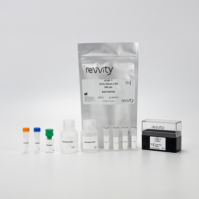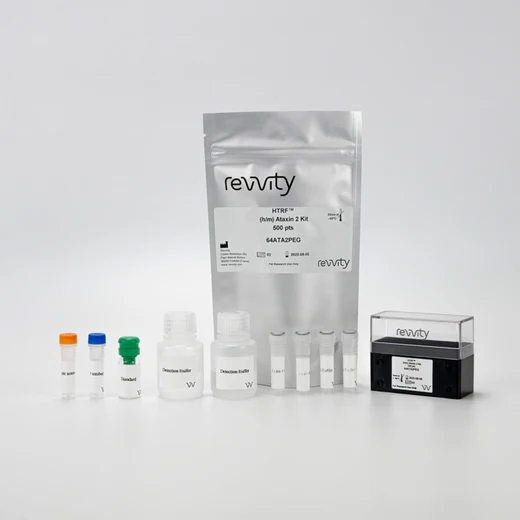

HTRF Human & Mouse Ataxin 2 Detection Kit, 500 Assay Points


HTRF Human & Mouse Ataxin 2 Detection Kit, 500 Assay Points


The HTRF Human and Mouse Ataxin 2 Detection Kit is designed for the quantitative measurement of Ataxin 2 in human and mouse cell lysates.
For research use only. Not for use in diagnostic procedures. All products to be used in accordance with applicable laws and regulations including without limitation, consumption and disposal requirements under European REACH regulations (EC 1907/2006).
| Feature | Specification |
|---|---|
| Application | Protein Quantification |
| Sample Volume | 16 µL |
The HTRF Human and Mouse Ataxin 2 Detection Kit is designed for the quantitative measurement of Ataxin 2 in human and mouse cell lysates.
For research use only. Not for use in diagnostic procedures. All products to be used in accordance with applicable laws and regulations including without limitation, consumption and disposal requirements under European REACH regulations (EC 1907/2006).


HTRF Human & Mouse Ataxin 2 Detection Kit, 500 Assay Points


HTRF Human & Mouse Ataxin 2 Detection Kit, 500 Assay Points


Product information
Overview
Ataxin 2 is an RNA-binding protein. In humans, Ataxin 2 causes neurodegeneration when carrying very long polyglutamine tract and drives disease progression in ALS. Current research in neurodegenerative and specifically ALS settings indicates there is a modulation of Ataxin 2 expression by TDP-43, which in turns sees its toxicity in cellular and animal models modified by Ataxin 2. The co-investigation of both proteins is a relevant topic for future therapeutic endeavours, with recent results indicating that lowered Ataxin 2 levels suppress TDP-43 aggregation.
Specifications
| Application |
Protein Quantification
|
|---|---|
| Brand |
HTRF
|
| Detection Modality |
HTRF
|
| Lysis Buffer Compatibility |
Lysis Buffer 1
Lysis Buffer 2
Lysis Buffer 3
Lysis Buffer 4
Lysis Buffer 5
|
| Product Group |
Kit
|
| Sample Volume |
16 µL
|
| Shipping Conditions |
Shipped in Dry Ice
|
| Target Class |
Biomarkers
|
| Target Species |
Human
Mouse
|
| Technology |
TR-FRET
|
| Unit Size |
500 Assay Points
|
Video gallery

HTRF Human & Mouse Ataxin 2 Detection Kit, 500 Assay Points

HTRF Human & Mouse Ataxin 2 Detection Kit, 500 Assay Points

How it works
Principle of the HTRF Human/Mouse Ataxin2 assay
The Human/Mouse Ataxin 2 assay is based on a TR-FRET sandwich immunoassay involving two specific antibodies, one labelled with Eu3+ cryptate (donor) and the other with d2 (acceptor). Both antibodies bind to Ataxin 2 (WT & mutant forms), and the donor-acceptor proximity enables a fluorescent TR-FRET signal. The intensity of the signal is directly proportional to the concentration of Ataxin 2 present in the sample (cell lysate or tissue lysate).

Human/Mouse Ataxin2 assay assay protocol
The Human/Mouse Ataxin 2 assay can be run in a 96- or 384-well low volume white detection plate (20 µL final). As described here, samples (cell/tissue lysates) or standards are dispensed directly into the assay plate for the detection of Ataxin 2 by HTRF® reagents. The antibodies labelled with HTRF fluorophores may be pre-mixed and added in a single dispensing step. No washing steps are needed. The protocol can be further miniaturized or upscaled by simply resizing each addition volume proportionally.

Assay details
Technical specifications of Human/Mouse Ataxin 2 kit
| Sample size | 5 µL |
|---|---|
| Final assay volume | 20 µL |
| Time to results | Overnight at RT |
| Detection limit (LOD) in lysis buffer #1 | 7 pg/mL |
| Dynamic range | 21 - 10,000 pg/mL |
| Species compatibility | Human and Mouse |

Analytical performance
Intra-assay precision table
| Sample | Mean [Ataxin2] (pg/mL) | CV |
|---|---|---|
| 1 | 100 | 10,0% |
| 2 | 640 | 3,6% |
| 3 | 1,600 | 6,7% |
| 4 | 4,000 | 6,2% |
| Mean CV | 6,6% |
| Sample | Mean [Ataxin2] (pg/mL) | CV |
|---|---|---|
| 1 | 100 | 6,1% |
| 2 | 640 | 1,7% |
| 3 | 1,600 | 0,5% |
| 4 | 4,000 | 3,8% |
|
|
Mean CV | 3,0% |
Each of the samples was measured in 3 independent experiments (3 separate days), and % CV was calculated for each sample.
Samples are cell lysate from WT Ataxin 2 transfected HEK293 cells.
Dilution table
| Dilution factor | [Ataxin2] Expected (pg/mL) | [Ataxin2] Measured (pg/mL) | Dilution recovery |
|---|---|---|---|
| Neat | - | 2,831 | 100% |
| 2 | 1,415 | 1,272 | 90% |
| 4 | 708 | 639 | 90% |
| 8 | 354 | 338 | 95% |
| 16 | 177 | 174 | 98% |
| Mean CV | 98% |
The excellent recovery percentages obtained from these experiments show the good dilution linearity of the assay. Samples are cell lysate from WT Ataxin2 transfected HEK293 cells, serially diluted in lysis buffer.
Assay validation
Staurosporine treatment on HTRF human/mouse Ataxin2 kit in ALS context
HeLa cells were plated at 100,000 cells/well in a 96-well plate and cultured at 37°C, 5% CO2 for 24 hours. The cell culture medium was removed, and 50µL of staurosporine solution in cell culture medium at 1µM were added. Treatment continued for 6 hours at 37°C, 5% CO2. Then the medium was removed and 50µL of lysis buffer were added. The plate was left under shaking for 30 minutes. Lysis followed or not the disaggregation protocol (from the TDP43 aggregation kit), and then 16µL of lysate were transfered into a 384 well plate for the detection, with 4µL of HTRF Ataxin 2 or HTRF Total TD43 detection reagent added to each well. Signals were recorded after overnight incubation.

Both Ataxin 2 and TDP-43 aggregation appear to increase following staurosporine treatment. This result is in line with the relationship between the two proteins as described in the literature. Current research in neurodegenerative and specifically ALS settings indicates there is a modulation of Ataxin 2 expression by TDP-43, which in turns has its toxicity in cellular and animal models modified by Ataxin 2. The co-investigation of these two proteins is a relevant topic for future therapeutic endeavours.
Selectivity of HTRF human/mouse Ataxin2 kit on KO cell line
HAP-1 KO cells were used to assess the selectivity of the assay. 2 KO cell lines from Horizon were available for Ataxin 2, one with 7pb depleted and the other with 67pb depleted. They were both tested.

100,000 cells of each cell line were plated in a 96-well plate and cultured at 37°C, 5% CO2 for 24 hours. Cell culture medium was removed, and 50µL of lysis buffer were added to the wells. The plate was shaken for 30 minutes and then lysates were transfered to a 384 well plate for the detection step. 4µL of HTRF Ataxin 2 detection reagent were added to each well, and HTRF signals were recorded after overnight incubation.
The results show normal detection of Ataxin 2 in the Ataxin3-KO cell line, and largely depleted to non-existent detection of Ataxin 2 in Ataxin 2-KO cell lines. This demonstrate the assay's good selectivity for Ataxin 2 compared to Ataxin3.
Sensitivity of HTRF human/mouse Ataxin2 kit on Ataxin2 mutant
To adress compatibility with different mutants, we designed 4 plasmids. One contained the WT Ataxin 2 protein sequence, and 3 contained increasing lengths of polyQ repeat motifs (30, 54, and 108 respectively).

Ataxin 2 HAP1 KO cell line (7pb depleted) was cultured in a T175 flask until confluency. Transfection was performed by incubating the cells with the different plasmids for 6 hours, before removing the transfection medium and adding cell culture medium for another 24 hours. Each flask was lysed with 3mL of lysis buffer, and then followed or not the disaggregation protocol. 16µL of each sample were transfered into a 384 well plate for the detection. 4µL of HTRF Ataxin 2 detection reagent were added to each well. The HTRF signals were recorded after overnight incubation.
Results show the assay recognizes mutant and WT Ataxin 2 proteins.
Detection of HTRF human/mouse Ataxin2 kit on Cell line
Neural cellular models were selected: SH-SY5Y (human) and Neuro2A (murine), HeLa cells (human) were also evaluated.

200,000, 100,000 or 50,000 cells of the different cell lines were plated in a 96-well plate and cultured at 37°C, 5% CO2 for 24 hours. The cell culture medium was removed and 50µL of lysis buffer was added to each well. The plate was then shaken for 30 minutes. Lysates were transfered to a 384 well plate for the detection step. 4µL of HTRF Ataxin 2 detection reagent were added to each well, and the HTRF signals were recorded after overnight incubation.
Simplified pathway
Ataxin 2 signaling pathway
Ataxin-2 binds to the mRNA of TDP-43, leading to an increase in mRNA stability and in turn to increased protein expression. This dysregulation affects the TDP-43 proteostasis and favors ALS disease onset.

TDP-43 (TAR DNA binding protein 43) is a DNA and RNA-binding protein which plays a crucial role in RNA metabolism. Mainly located in the nucleus, TDP-43 shuttles between the nucleus and the cytoplasm. In pathological conditions, including stress or mutation, there is a mislocalization of TDP-43 in the cytoplasm. It can be cleaved into C terminal fragments, thus creating an accumulation of ubiquitinated and hyperphosphorylated toxic aggregates, and then insoluble inclusion bodies. Mislocalized soluble TDP-43 becomes ubiquitined to be degraded by the proteasome, whereas autophagy tries to remove insoluble aggregates when it is still possible.
Resources
Are you looking for resources, click on the resource type to explore further.
Discover the versatility and precision of Homogeneous Time-Resolved Fluorescence (HTRF) technology. Our HTRF portfolio offers a...


How can we help you?
We are here to answer your questions.


