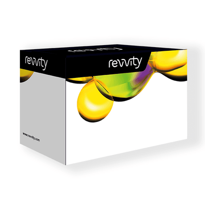

HTRF Human Total Aurora A Detection Kit, 500 Assay Points


HTRF Human Total Aurora A Detection Kit, 500 Assay Points


The Total Aurora A kit is designed to monitor the expression level of cellular human Aurora A, and can be used as a normalization assay for the Phospho-human Aurora A Thr288 Detection Kit.
For research use only. Not for use in diagnostic procedures. All products to be used in accordance with applicable laws and regulations including without limitation, consumption and disposal requirements under European REACH regulations (EC 1907/2006).
| Feature | Specification |
|---|---|
| Application | Cell Signaling |
| Sample Volume | 16 µL |
The Total Aurora A kit is designed to monitor the expression level of cellular human Aurora A, and can be used as a normalization assay for the Phospho-human Aurora A Thr288 Detection Kit.
For research use only. Not for use in diagnostic procedures. All products to be used in accordance with applicable laws and regulations including without limitation, consumption and disposal requirements under European REACH regulations (EC 1907/2006).


HTRF Human Total Aurora A Detection Kit, 500 Assay Points


HTRF Human Total Aurora A Detection Kit, 500 Assay Points


Product information
Overview
This HTRF cell-based assay conveniently and accurately detects human Aurora A protein (related to the AURKA gene) ubiquitously expressed in eukaryotes. Aurora A belongs to the serine/threonine Aurora kinases family (including Aurora B and Aurora C), essential for cell division via mitosis regulation and especially the process of chromosomal segregation. It is activated through the autophosphorylation of Threonine 288 residue mainly occurring from the late S-Phase through the M phase during the cellular division cycle or DNA damage response. The overexpression of the Aurora kinases in a wide range of cancers (Leukemia, Colon Cancer, Prostate Cancer, and Breast Cancer) makes them interesting candidates for the development of Aurora kinase inhibitors (AKIs).
Specifications
| Application |
Cell Signaling
|
|---|---|
| Brand |
HTRF
|
| Detection Modality |
HTRF
|
| Lysis Buffer Compatibility |
Lysis Buffer 1
Lysis Buffer 4
|
| Molecular Modification |
Total
|
| Product Group |
Kit
|
| Sample Volume |
16 µL
|
| Shipping Conditions |
Shipped in Dry Ice
|
| Target Class |
Phosphoproteins
|
| Target Species |
Human
|
| Technology |
TR-FRET
|
| Unit Size |
500 Assay Points
|
Video gallery

HTRF Human Total Aurora A Detection Kit, 500 Assay Points

HTRF Human Total Aurora A Detection Kit, 500 Assay Points

How it works
Total Aurora A assay principle
The Total-Aurora A assay quantifies the expression level of Aurora A in a cell lysate. Unlike Western Blot, the assay is entirely plate-based and does not require gels, electrophoresis, or transfer. The Total-Aurora A assay uses two labeled antibodies: one coupled to a donor fluorophore, the other to an acceptor. Both antibodies are highly specific for a distinct epitope on the protein. In presence of Aurora A in a cell extract, the addition of these conjugates brings the donor fluorophore into close proximity with the acceptor and thereby generates a FRET signal. Its intensity is directly proportional to the concentration of the protein present in the sample, and provides a means of assessing the protein’s expression under a no-wash assay format.

Total-Aurora A 2-plate assay protocol
The 2-plate protocol involves culturing cells in a 96-well plate before lysis, then transferring the lysates into a 384-well low volume detection plate before the addition of Total Aurora A HTRF detection reagents. This protocol enables the cells' viability and confluence to be monitored.

Total-Aurora A 1-plate assay protocol
Detection of total Aurora A with HTRF reagents can be performed in a single plate used for culturing, stimulation, and lysis. No washing steps are required. This HTS designed protocol enables miniaturization while maintaining robust HTRF quality.

Assay validation
Activation of Total and Phospho-Aurora A (Thr288) measured on HeLa cells
HeLa cells (Immortalized Human cervical cancer cell line) were seeded in a 96-well culture-treated plate at 100,000 cells/well for 24 hours in complete culture medium for adhesion. After cell culture medium removal, the cells were treated with increasing concentrations of Nocodazole for 20h at 37° C, 5% CO2. After overnight treatment (20h), the cell culture medium was removed and 50 µl of supplemented Lysis Buffer#1 (1X) were dispensed into each well for 30 min at RT under gentle shaking. After cell lysis, 16 µL of lysates were transferred into a 384-well low volume white microplate and 4 µL of the HTRF Total Aurora A or Phospho-Aurora A (Thr288) detection antibodies were added. The HTRF signal was recorded after an overnight incubation.
Nocodazole is a microtubule destabilizer inducing a G2/M cell cycle arrest. It has been demonstrated that Nocodazole-treated cells show higher Aurora protein expression levels to overcome cell cycle arrest [1].
As expected, the results obtained show an increased level of Total and Thr288 Phosphorylated Aurora A proteins upon treatment with Nocodazole.
[1] : Jiang et al. Oncogene,2003

Inhibitor characterization with Phospho-Aurora A (Thr288) and Total Aurora A on HeLa cells


Total and phospho Aurora A on HeLa cells treated with Alisertib
HeLa cells (Immortalized Human cervical cancer cell line) were seeded in a 96-well culture-treated plate at 100,000 cells/well for 24 hours in complete culture medium for adhesion. After cell culture medium removal, they were treated with increasing concentrations of inhibitors ( MLN8054, Tozasertib, or Alisertib) co-incubated with a fixed concentration of Nocodazole at 200 nM for 20h at 37° C, 5% CO2, in cell culture medium containing 5% SVF. After overnight treatment, the cell culture medium was removed and 50 µl of supplemented Lysis Buffer#1 (1X) were dispensed into each well for 30 min at RT under gentle shaking. After cell lysis, 16 µL of lysates were transferred into a 384-well low volume white microplate and 4 µL of the HTRF Total Aurora A or Phospho-Aurora A (Thr288) detection antibodies were added. The HTRF signal was recorded after an overnight incubation.
The 3 compounds have been described in the literature for their inhibitory effect on phosphorylated Aurora A (thr288), reaching 98% maximum inhibition, without impacting the total protein level [2,3,4,5].
As expected, the results obtained show a clear dose-dependent inhibition of the Aurora A phosphorylation at Thr288 upon treatment with MLN8054, Tozasertib, or Alisertib, while the Aurora A protein expression level remains constant.
[2] : Manfredi et al. PNAS,2006
[3] : Sells et al. ACS Med.Chem.Lett.,2015
[4] : Tyler et al. Cell cycle,2007
[5] : Qi et al. Leuk Res,2013

Specificity and selectivity of HTRF Total Aurora A assay using gene silencing (SiRNA)
HeLa cells were treated with 25 nM of SMARTPool ON-TARGETplus siRNA (Horizon) specifically targeting Aurora A (#L-003545-01-0005), Aurora B (# L-003326-00-0005), and Aurora C (#L-019573-00-0005), or with a non-targeting siRNA (# D-001810-10-20, included as control), in a 96-well plate (25,000 cells/well) under 100 µL for 24H. After cell culture medium removal, cells were stimulated by adding 300 nM Nocodazole in complete cell culture medium for an additional 24h-incubation at 37°C, 5% CO2. After medium removal, cells were lysed with 50 µL lysis buffer #1 (1X) for 30 min at RT under gentle shaking, and 16 µL of lysates were transferred into a low volume white microplate before the addition of 4 µL of premixed HTRF Total Aurora A detection antibodies. The HTRF signal was recorded after an overnight incubation at RT.
Cell treatment with Aurora A siRNA led to a significant downregulation of Aurora A with a 44% signal decrease compared to the cells transfected with the non-targeting siRNA.
Despite high homology between the 3 family proteins, no decrease in signal was observed for cells treated with Aurora B or Aurora C siRNA, demonstrating the specificity and selectivity of the kit.

Validation on various Human cell lines
Immortalized human cell lines HepG2 (liver cancer), HeLa (Cervix cancer), HEK293, and HCT116 (Colon Cancer) were plated in 96-well culture plates at a density of 25,000 or 100,000 cells /well and incubated for 24 hours at 37°C, 5% CO2. After cell culture medium removal, cells were stimulated by adding 300 nM Nocodazole in complete cell culture medium for 20H at 37°C, 5% CO2. Cells were lysed after medium removal with 50 µL of supplemented lysis buffer #1 (1X), for 30 min at RT under gentle shaking.
The Aurora A protein expression level was assessed with the HTRF Total Aurora A kit. Briefly, 16 µL of cell lysate were transferred into a low volume white microplate, followed by 4 µL of premixed HTRF detection reagents. The HTRF signal was recorded after an overnight incubation at RT.
Aurora A protein is well-detected in all the tested human cell lines at different levels. For a determined cellular model, cell density optimization is mandatory to be within the dynamic range of the kit. Note that HepG2, which expresses higher Aurora A protein levels, must be used at a lower cell density to be within the dynamic range (25,000 cells/well or below) compared to HeLa, HEK293, or HCT116 ( 25,000 to 100,000 cells/well).
The HTRF Total Aurora A assay efficiently detects endogenous Aurora A protein in various human cellular models expressing different levels of the protein.

HTRF Total Aurora A assay compared to Western Blot
HeLa cells were cultured in a T175 flask in complete culture medium at 37°C, 5% CO2. After 24H incubation, the cells were first stimulated with Nocodazole 300 nM for 20H, 37°C, 5% CO2, then lysed with 3 mL of supplemented lysis buffer #1 (1X) for 30 minutes at RT under gentle shaking.
Serial dilutions of the cell lysate were performed using supplemented lysis buffer, and 16 µL of each dilution were transferred into a low volume white microplate before the addition of 4 µL of HTRF Total Aurora A detection reagents. Equal amounts of lysates were used for a side by side comparison between HTRF and Western Blot.
A side by side comparison of Western Blot and HTRF demonstrates that the HTRF assay is 4-fold more sensitive than the Western Blot, at least under these experimental conditions.

Simplified pathway
Simplified Aurora A signaling pathway
Aurora kinases, a family of serine/threonine kinases consisting of Aurora A (AURKA), Aurora B (AURKB), and Aurora C (AURKC), are essential kinases for cell division via mitosis regulation, especially in the process of chromosomal segregation.
Aurora A activation, through the autophosphorylation of Threonine 288 residue on its activation loop, mainly occurs from the late S-Phase through the M phase during the cellular division cycle or DNA damage response. Several co-factors, including microtubule associated protein TPX2 and INCENP, are required for this activation. Aurora A phosphorylates several targets such as PLK1 (in association with its cofactor Bora) to activate PLK1 cascades controlling key aspects of mitosis and spindle assembly, or activating the recovery pathway in response to DNA-damage . Aurora A is inactivated through dephosphorylation of Thr288 by protein phosphatase 1 (PP1). The overexpression of the Aurora kinases in a wide range of cancers (Leukemia, Colon Cancer, Prostate Cancer and Breast Cancer) make them interesting candidates for the development of Aurora kinase inhibitors (AKIs).

Resources
Are you looking for resources, click on the resource type to explore further.
This guide provides you an overview of HTRF applications in several therapeutic areas.


How can we help you?
We are here to answer your questions.


