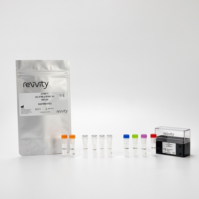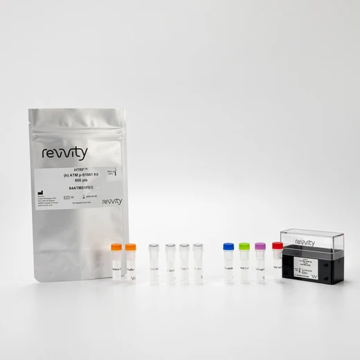

HTRF Human Phospho-ATM (Ser1981) Detection Kit, 10,000 Assay Points


HTRF Human Phospho-ATM (Ser1981) Detection Kit, 10,000 Assay Points


This HTRF kit enables the cell-based quantitative detection of ATM phosphorylation at Ser1981, which is activated upon DNA damage. This kit enables optimal investigation of the ATM/CHK2 pathway, including selective inhibitors.
For research use only. Not for use in diagnostic procedures. All products to be used in accordance with applicable laws and regulations including without limitation, consumption and disposal requirements under European REACH regulations (EC 1907/2006).
| Feature | Specification |
|---|---|
| Application | Cell Signaling |
| Sample Volume | 16 µL |
This HTRF kit enables the cell-based quantitative detection of ATM phosphorylation at Ser1981, which is activated upon DNA damage. This kit enables optimal investigation of the ATM/CHK2 pathway, including selective inhibitors.
For research use only. Not for use in diagnostic procedures. All products to be used in accordance with applicable laws and regulations including without limitation, consumption and disposal requirements under European REACH regulations (EC 1907/2006).


HTRF Human Phospho-ATM (Ser1981) Detection Kit, 10,000 Assay Points


HTRF Human Phospho-ATM (Ser1981) Detection Kit, 10,000 Assay Points


Product information
Overview
This HTRF cell-based assay enables the rapid, quantitative detection of ATM phosphorylated at Serine 1981, as a readout of the ATM/CHK2 signaling pathway upon a DNA damage response (DDR). This kit is complementary to the phospho CHK2 Thr68 for studies of the ATM-CHK2 pathway.
In response to DNA damage, such as Double Strand Breaks (DSBs), the ATM€“Chk2 pathway and replication checkpoint responses are activated. These mediate G1/S checkpoints to arrest cell cycle progression and allow extra time for DNA repair.
In response to DNA DSBs, ATM autophosphorylates to generate the active monomeric kinase. Activation of ATM results in the phosphorylation of a diverse array of downstream targets such as H2A histone family member X, H2Ax, or the checkpoint kinase Chk2 and its downstream effectors.
Specifications
| Application |
Cell Signaling
|
|---|---|
| Brand |
HTRF
|
| Detection Modality |
HTRF
|
| Lysis Buffer Compatibility |
Lysis Buffer 1
Lysis Buffer 4
|
| Molecular Modification |
Phosphorylation
|
| Product Group |
Kit
|
| Sample Volume |
16 µL
|
| Shipping Conditions |
Shipped in Dry Ice
|
| Target Class |
Phosphoproteins
|
| Target Species |
Human
|
| Technology |
TR-FRET
|
| Unit Size |
10,000 Assay Points
|
Video gallery

HTRF Human Phospho-ATM (Ser1981) Detection Kit, 10,000 Assay Points

HTRF Human Phospho-ATM (Ser1981) Detection Kit, 10,000 Assay Points

How it works
HTRF phospho ATM assay principle
The Phospho-ATM (Ser1981) assay measures ATM when phosphorylated at Serine 1981. Unlike Western Blot, the assay is entirely plate-based and does not require gels, electrophoresis, or transfer.
The Phospho-ATM (Ser 1981) assay uses 2 labeled antibodies: one with a donor fluorophore, the other with an acceptor. The first antibody was selected for its specific binding to the phosphorylated motif on the protein, and the second for its ability to recognize the protein independently of its phosphorylation state. Protein phosphorylation enables an immune-complex formation involving the two labeled antibodies and which brings the donor fluorophore into close proximity to the acceptor, thereby generating a FRET signal. Its intensity is directly proportional to the concentration of phosphorylated protein present in the sample, and provides a means of assessing the protein’s phosphorylation state under a no-wash assay format.

HTRF Phospho-ATM Ser 1981 2-plate assay protocol
The 2 plate protocol involves culturing cells in a 96-well plate before lysis, then transferring lysates to a 384-well low volume detection plate before the addition of the phospho-ATM ser 1981 HTRF detection reagents.
This protocol enables the cells' viability and confluence to be monitored.

HTRF Phospho-ATM Ser 1981 one-plate assay protocol
Detection of phospho ATM Ser 1981 with HTRF reagents can be performed in a single plate used for culturing, stimulation, and lysis. No washing steps are required.
This HTS-designed protocol enables miniaturization while maintaining robust HTRF quality.

Assay validation
Neocarzinostatin effect on total and phospho Ser 1981 ATM assay
Human HEK293 cells were plated in a 96-well culture-treated plate (100,000 cells/well) in complete culture medium, and incubated overnight at 37°C, 5% CO2. Cells were treated with a dose-response of neocarzinostatin 2h at 37 °C, 5% CO2. They were then lysed with 50 µl of supplemented lysis buffer #1 (1X) for 30 min at RT under gentle shaking. After cell lysis, 16 µL of lysate were transferred into a 384-well low volume white microplate and 4 µL of the HTRF Phospho ATM (Ser 1981) or Total-ATM detection reagents were added. The HTRF signal was recorded after an overnight incubation at room temperature.
As expected, neocarzinostatin induced single and double strand DNA damage, leading to a dose-dependent increase in ATM phosphorylation, without any effect on the expression level of the ATM total protein.

Effect of compounds inducing DNA damage on ATM phosphorylation (Ser1981) and total protein
Human HEK293 cells were plated in a 96-well culture-treated plate (100,000 cells/well) in complete culture medium, and incubated overnight at 37°C, 5% CO2. Cells were treated with a dose-response of neocarzinostatin, hydroxyurea, doxorubicin, and etoposide, for 2h at 37 °C, 5% CO2. The medium was then removed, and the cells were lysed with 50 µl of supplemented lysis buffer #1 (1X) for 30 min at RT under gentle shaking. After cell lysis, 16 µL of lysate were transferred into a 384-well low volume white microplate and 4 µL of the HTRF Phospho ATM (Ser1981) or Total-ATM detection reagents were added. The HTRF signal was recorded after an overnight incubation at room temperature.
The different compounds showed different responses. Neocarzinostatin, doxorubicin, and etoposide are known to induce double strand breaks (DSB), and here led to phosphorylation of ATM. On the other hand hydroxyurea, which preferably induces single strand breaks (SSBs), displayed a partial ATM phosphorylation with weak potency. The EC50 of neocarzinostatin, doxorubicin, etoposide, and hydroxyurea were evaluated at 0.2 µM, 1.4 µM, 0.9 µM, and 0.3 mM respectively.
Moreover, the EC80 of neocarzinostatin was evaluated at 1 µM, and this concentration was used to assess inhibitors of the ATM/CHK2 Pathway.
None of the 4 compounds affected the expression level of the ATM total protein.


Effect of ATR/CHK1 or ATM/CHK2 pathway inhibitors on HTRF Phospho Ser1981 and Total ATM kits
Human HEK293 cells were plated in a 96-well culture-treated plate (100,000 cells/well) in complete culture medium, and incubated overnight at 37°C, 5% CO2. Cells were treated with a dose-response of 3 inhibitors of the ATR or ATM pathways for 2h at 37 °C, 5% CO2. The cells were then treated with 1 µM of neocarzinostatin (EC80) for another 2h at 37 °C, 5% CO2. The medium was removed, and the cells were then lysed with 50 µl of supplemented lysis buffer #1 (1X) for 30 min at RT under gentle shaking. After cell lysis, 16 µL of lysate were transferred into a 384-well low volume white microplate and 4 µL of the HTRF Phospho ATM Ser 1981 or Total-ATM detection reagents were added. The HTRF signal was recorded after an overnight incubation at room temperature.
Caffeine is known to be a mild ATR/ATM pathway inhibitor, UCN-1 is a potent CHK1 inhibitor, and KU55933 is a selective ATM pathway inhibitor. As expected, UCN-1 had no effect on ATM phosphorylation. Caffeine showed a decrease in ATM phosphorylation with weak potency. KU55933 allowed a full inhibition of ATM phosphorylation with higher potency.
These 3 compounds did not affect the expression level of the ATM total protein.


Simplified pathway
ATM/CHK2 and ATR/CHK1 signaling pathways
DNA double-strand breaks (DSBs) or single-strand breaks (SSBs) are among the most deleterious lesions that threaten genome integrity. They can be induced by the effect of cellular metabolites or by DNA-damaging agents such as genotoxic compounds, chemotherapeutic agents, ultraviolet (UV) irradiation, or ionizing radiation.
DNA damage response (DDR) is mainly controlled by ataxia telangiectasia mutated (ATM) and by ataxia telangiectasia and Rad3-related (ATR), two members of the phosphoinositide 3-kinase (PI3K)-related kinase (PIKK) protein kinase family.
In response to DNA damage (DSBs), ATM–Chk2 pathway and replication checkpoint responses are activated, mediating G1/S checkpoints to arrest cell cycle progression and allow extra time for DNA repair.
Reacting to DNA DSBs, ATM autophosphorylates to generate the active monomeric kinase. Activation of ATM results in the phosphorylation of a diverse array of downstream targets such as H2A histone family member X, H2Ax, or the checkpoint kinase Chk2 and its downstream effectors.
The isolation of new small molecule inhibitors of ATM, and the design and validation of novel strategies of checkpoint modulation, combined with the traditional radiation and chemotherapy modalities, hold promise for improved treatment of cancers.

Resources
Are you looking for resources, click on the resource type to explore further.
This document includes detailed tables listing HTRF™, AlphaLISA™ SureFire® Ultra™, and Alpha SureFire® Ultra™ Multiplex assays...


How can we help you?
We are here to answer your questions.


