


US
Revvity Sites Globally
Select your location.
*e-commerce not available for this region.

HTRF Human Phospho-EGFR (Tyr1068) Detection Kit, 500 Assay Points


HTRF Human Phospho-EGFR (Tyr1068) Detection Kit, 500 Assay Points


Shipping box for Revvity reagent kits


The phospho-EGFR (Tyr1068) kit is designed for the cell-based quantitative detection of EGFR phosphorylation.
For research use only. Not for use in diagnostic procedures. All products to be used in accordance with applicable laws and regulations including without limitation, consumption and disposal requirements under European REACH regulations (EC 1907/2006).
| Feature | Specification |
|---|---|
| Application | Cell Signaling |
| Sample Volume | 16 µL |
The phospho-EGFR (Tyr1068) kit is designed for the cell-based quantitative detection of EGFR phosphorylation.
For research use only. Not for use in diagnostic procedures. All products to be used in accordance with applicable laws and regulations including without limitation, consumption and disposal requirements under European REACH regulations (EC 1907/2006).
Product Variants
Unit Size: 500 Assay Points
Part #:
64EG1PEG
List Price
USD 2,147.00
Unit Size: 10,000 Assay Points
Part #:
64EG1PEH
List Price
USD 12,490.00
HTRF Human Phospho-EGFR (Tyr1068) Detection Kit, 500 Assay Points
Shipping box for Revvity reagent kits
HTRF Human Phospho-EGFR (Tyr1068) Detection Kit, 500 Assay Points
Product information
Overview
The phospho-EGFR assay kit is designed for the cell-based quantitative detection of Tyr1068 phosphorylation on EGFR. Mutations that lead to an overexpression or hyperactivity of EGFR, also known as ErbB1 or HER1, are associated with a number of cancers, such as lung, colorectal, prostate, glioblastoma cancer, etc. This makes EGFR a key target for anti-cancer therapy-related immunoassay.
Specifications
| Application |
Cell Signaling
|
|---|---|
| Brand |
HTRF
|
| Detection Modality |
HTRF
|
| Molecular Modification |
Phosphorylation
|
| Product Group |
Kit
|
| Sample Volume |
16 µL
|
| Shipping Conditions |
Shipped in Dry Ice
|
| Target Class |
Phosphoproteins
|
| Target Species |
Human
|
| Technology |
TR-FRET
|
| Therapeutic Area |
Oncology & Inflammation
|
| Unit Size |
500 Assay Points
|
Video gallery
HTRF Human Phospho-EGFR (Tyr1068) Detection Kit, 500 Assay Points
HTRF Human Phospho-EGFR (Tyr1068) Detection Kit, 500 Assay Points
Citations
How it works
Phospho-EGFR (Tyr1068) assay principle
The Phospho-EGFR (Tyr1068) assay measures EGFR when phosphorylated at Tyr1068. Contrary to Western Blot, the assay is entirely plate-based and does not require gels, electrophoresis or transfer. The Phospho-EGFR (Tyr1068) assay uses 2 labeled antibodies: one with a donor fluorophore, the other one with an acceptor. The first antibody is selected for its specific binding to the phosphorylated motif on the protein, the second for its ability to recognize the protein independent of its phosphorylation state. Protein phosphorylation enables an immune-complex formation involving both labeled antibodies and which brings the donor fluorophore into close proximity to the acceptor, thereby generating a FRET signal. Its intensity is directly proportional to the concentration of phosphorylated protein present in the sample, and provides a means of assessing the proteins phosphorylation state under a no-wash assay format.
Phospho-EGFR (Tyr1068) 2-plate assay protocol
The 2 plate protocol involves culturing cells in a 96-well plate before lysis then transferring lysates to a 384-well low volume detection plate before adding Phospho-EGFR (Tyr1068) HTRF detection reagents. This protocol enables the cells' viability and confluence to be monitored.

Phospho-EGFR (Tyr1068) 1-plate assay protocol
Detection of Phosphorylated EGFR (Tyr1068) with HTRF reagents can be performed in a single plate used for culturing, stimulation and lysis. No washing steps are required. This HTS designed protocol enables miniaturization while maintaining robust HTRF quality.

Assay validation
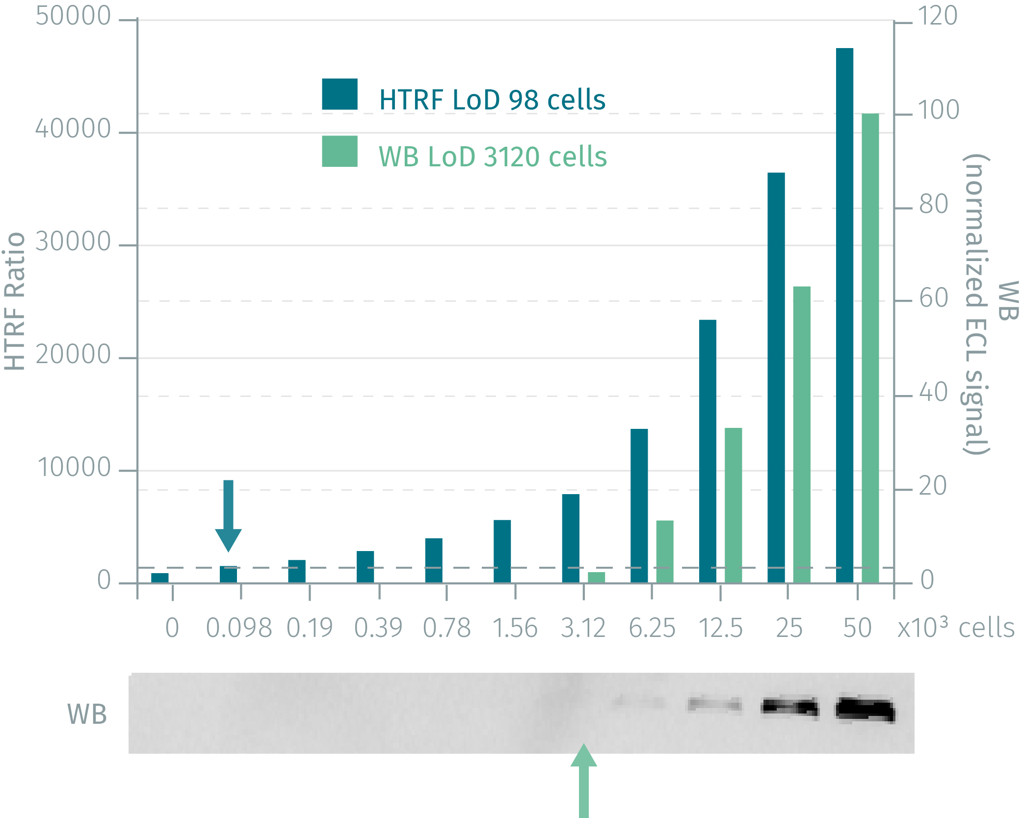
HTRF phospho-EGFR (Tyr1068) assay compared to WB
Human A431 cells were cultured for 48 h and stimulated with 100 EGF for 10 min. Following lysis, soluble fractions were collected via centrifugation. Serial dilutions of the cell lysate were performed in the supplemented lysis buffer and 16 µL of each dilution were dispensed and analyzed side-by-side by Western-blot and by HTRF. By using HTRF phospho-EGFR (Tyr1068) only 100 cells are sufficient for minimal signal detection while 3,120 cells are needed for a Western Blot signal. The HTRF phospho-EGFR assay is at least 30-fold more sensitive than the Western Blot and shows optimal correlation.

Phospho-EGFR modulation in several human tumor cell lines
Human BxPC3 (Figure 1;green), SKOV3 (Figure 2;purple), CRC1 (Figure 3) and A431 (Figure 4;pink) cells were plated in 96 well plate and incubated for 24 h, at 37 °C-5% Co2. After 10 min incubation with increasing concentrations of agonist, medium was removed and cells were lysed with 50 µL of Lysis buffer for 30min at RT under gentle shaking.16 µL of lysate was transfered into a 384 sv white microplate and 4 µL of the HTRF phospho-EGFR detection reagents were added. HTRF signal was recorded after an overnight incubation.


Inhibition measured with Phospho (tyr1068) & total EGFR kits


Simplified pathway
EGFR Epidermal growth factor receptor signaling Simplified Pathway
EGFR is a receptor tyrosine kinase that belongs to the ErbB family. Binding of EGFR ligands drive receptor homodimerization or hetero-dimerization leading to the activation of the EGFR tyrosine kinase domain and specific tyrosine residues.
The phosphorylated tyrosine residues then provide docking sites for a variety of factors that induce downstream activation of several signal transduction cascades.
The transmitted signals from the EGF receptor to the nucleus lead to the regulation of various biological functions such as cell proliferation, differentiation, survival, adhesion, migration & angiogenesis.
Mutations that lead to overexpression or hyperactivity of EGFR are associated with a number of cancers making EGFR a key target for anti-cancer therapies.

Resources
Are you looking for resources, click on the resource type to explore further.
Brochure
HTRF assays and reagents catalog
Discover the versatility and precision of Homogeneous Time-Resolved Fluorescence (HTRF) technology. Our HTRF portfolio offers a...
Guide
HTRF solutions, guide to major applications
This guide provides you an overview of HTRF applications in several therapeutic areas.
SDS, COAs, Manuals and more
Are you looking for technical documents related to the product? We have categorized them in dedicated sections below. Explore now.
Safety data sheet
-
LanguageEnglishCountryUnited States
-
LanguageFrenchCountryFrance
-
LanguageGermanCountryGermany
-
LanguageGreekCountryGreece
-
LanguageItalianCountryItaly
-
LanguageSpanishCountrySpain
-
LanguageEnglishCountryUnited Kingdom
+ Show more
Certificate of analysis
-
Lot number13LLot dateFebruary 21, 2025
-
Lot number13JLot dateFebruary 21, 2025
-
Lot number13MLot dateFebruary 21, 2025
-
Lot number13HLot dateFebruary 21, 2025
-
Lot number13KLot dateFebruary 21, 2025
-
Lot number13GLot dateFebruary 21, 2025
-
Lot number13FLot dateNovember 20, 2024
+ Show more
IFUs, Manuals & more
-
Resource typeManualLanguageEnglishCountry-


How can we help you?
We are here to answer your questions.

