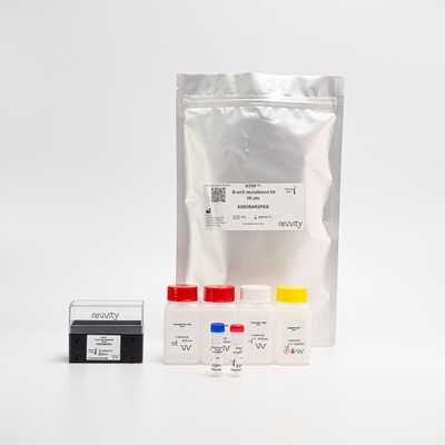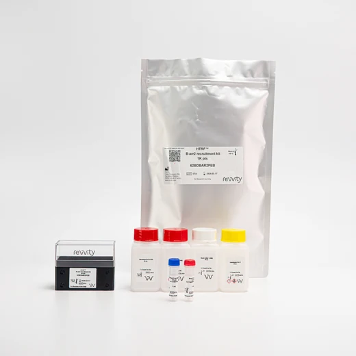

HTRF β-arrestin 2 Recruitment Detection Kit, 1,000 Assay Points


HTRF β-arrestin 2 Recruitment Detection Kit, 1,000 Assay Points


The B-arr2 recruitment cellular assay monitors the recruitment of B-arrestin 2 and AP2 following GPCR activation.
For research use only. Not for use in diagnostic procedures. All products to be used in accordance with applicable laws and regulations including without limitation, consumption and disposal requirements under European REACH regulations (EC 1907/2006).
| Feature | Specification |
|---|---|
| Application | Second Messenger Detection |
| Sample Volume | 200 µL |
The B-arr2 recruitment cellular assay monitors the recruitment of B-arrestin 2 and AP2 following GPCR activation.
For research use only. Not for use in diagnostic procedures. All products to be used in accordance with applicable laws and regulations including without limitation, consumption and disposal requirements under European REACH regulations (EC 1907/2006).


HTRF β-arrestin 2 Recruitment Detection Kit, 1,000 Assay Points


HTRF β-arrestin 2 Recruitment Detection Kit, 1,000 Assay Points


Product information
Overview
The B-arr2 recruitment cellular assay monitors the recruitment of Beta-arrestin 2 and AP2 through GPCR activation. The B-arr2 recruitment kit is used to characterize the effect of compounds on the Beta-arrestin signaling pathway, by monitoring the interaction between endogenous B-arrestin 2 and AP-2 as its partner.
It is known from the literature that pharmacological investigations on this interaction are relevant when investigating the Beta-arrestin signaling pathway in a GPCR context.
Specifications
| Application |
Second Messenger Detection
|
|---|---|
| Brand |
HTRF
|
| Detection Modality |
HTRF
|
| Product Group |
Kit
|
| Sample Volume |
200 µL
|
| Shipping Conditions |
Shipped in Dry Ice
|
| Target Class |
GPCR
|
| Technology |
TR-FRET
|
| Therapeutic Area |
Cardiovascular
Infectious Diseases
Inflammation
Metabolism/Diabetes
NASH/Fibrosis
Neuroscience
Oncology & Inflammation
Rare Diseases
|
| Unit Size |
1,000 Assay Points
|
Video gallery

HTRF β-arrestin 2 Recruitment Detection Kit, 1,000 Assay Points

HTRF β-arrestin 2 Recruitment Detection Kit, 1,000 Assay Points

Citations
How it works
B-arr2 recruitment kit assay principle
The B-arr2 recruitment assay monitors the recruitment of B-arrestin 2 mediated by GPCR activation in cells. Revvity's B-arr2 recruitment kit is highly sensitive, enabling the pharmacological characterization of compounds involving endogenous B-arrestin 2 and AP2 proteins. The kit uses two labeled antibodies, one coupled to a donor fluorophore, and the other to an acceptor. Both antibodies are highly specific for B-arr2 protein and AP2. In presence of B-arr2/AP2 interaction in cells, the addition of these conjugates brings the donor fluorophore into close proximity with the acceptor, and thereby generates a FRET signal. Its intensity is directly proportional to the number of B-arrestin 2/AP2 complexes present in the sample.

B-arr2 recruitment assay protocol (option 1)
Detection of B-arr2 recruitment with HTRF reagents can be performed in a single plate used for culturing, stimulation, stabilization, and detection. Three washes are required after the stabilization step. This specific protocol enables detection with non-lysed cells and endogenous proteins after ON incubation at room temperature, while maintaining robust HTRF quality.

B-arr2 recruitment assay protocol (option 2)
Detection of B-arr2 recruitment with HTRF reagents can be performed in a single plate used for culturing, stimulation, stabilization and detection. Three washes are required after the stabilization step. This specific protocol enables detection with non-lysed cells and endogenous proteins, while maintaining robust HTRF quality after ON incubation at room temperature.
Option 2 of the B-arr2 recruitment assay protocol enables improvement of S/B by removing 80µL of detection reagents after ON incubation at room temperature.

Assay validation
Illustrations with agonist and antagonist characterization on two GPCRs, as models
Tag-lite SNAP-Beta2R and SNAP-V1AR HEK293 cells were plated at 100,000 cells/well in a 96-well plate, and incubated for 24h at 37°C, 5% CO2. This was followed by treatment for 5 minutes (Vasopressin V1A receptor) and 30 minutes (Beta2 adrenergic receptor) at room temperature, with increasing concentrations of respectively Vasopressin (V1AR) and Isoproterenol (Beta2R) diluted in Stimulation buffer 4. The medium was next removed. Cells were stabilized with 30 µL of Stabilization buffer 1 for 15 minutes at room temperature, then washed 3 times with 100µl of Wash buffer 1. Finally, 100µl of the B-arr2 recruitment detection reagents were added. The HTRF signal was recorded after an overnight incubation at room temperature.
Following a similar protocol, antagonist compounds were characterized by dispensing increasing concentrations of SR 49059 (V1AR) and ICI-118,551 (Beta2R) for 30minutes at room temperature before adding agonist compounds at 15nM (Vasopressin) and 40nM (Isoproterenol).


Ranking of pharmacological compounds on Beta2 adrenergic receptor in HEK293 cells
Tag-lite SNAP-Beta2R HEK293 cells were plated at 100,000 cells/well in a 96-well plate, and incubated for 24h at 37°C, 5% CO2. After treatment for 30 minutes at room temperature with increasing concentrations of a panel of agonist compounds diluted in Stimulation buffer 4, the medium was removed. The cells were stabilized with 30 µL of Stabilization buffer 1 for 15 minutes at room temperature, then washed 3 times with 100µl of Wash buffer 1. Finally, 100µl of the B-arr2 recruitment detection reagents were added. The HTRF signal was recorded after an overnight incubation at room temperature.
Following a similar protocol, a panel of antagonist compounds were characterized by dispensing increasing concentrations of antagonists for 30 minutes at room temperature before adding Isoproterenol at 30nM.


Comparison of detection with assay protocol option 1 vs option 2
Tag-lite SNAP-GLP1R HEK293 cells were plated at 100,000 cells/well in a 96-well plate, and incubated for 24h at 37°C, 5% CO2. After treatment for 30 minutes at room temperature with increasing concentrations of Exendin-4 diluted in Stimulation buffer 4, the medium was removed. The cells were stabilized with 30 µL of Stabilization buffer 1 for 15 minutes at room temperature, then washed 3-times with 100µl of Wash buffer 1. Finally, 100µl of the B-arr2 recruitment detection reagents were added. The HTRF signal was recorded after an overnight incubation at room temperature (assay protocol option 1).
An additional step was performed after the overnight incubation, with the removal of 80µL of detection reagents (assay protocol option 2). The improved HTRF signal was recorded by using the same plate reader settings as for assay protocol option 1.

Simplified pathway
Simplified GPCR signaling pathway
B-arrestins play central roles in the GPCR signaling pathways by regulating agonist-mediated GPCR signaling. Their actions include mediating both receptor desensitization and resensitization processes, acting as a signaling scaffold for MAPK pathways such as MAPK1/3 or AKT1, and participating in the recruitment of the ubiquitin-protein ligase to the receptors. Beta-arrestins function as multivalent adapter proteins that can switch the GPCR from a G-protein signaling mode (that transmits short-lived signals from the plasma membrane) to a beta-arrestin signaling mode that transmits a distinct set of signals, initiated as the receptor internalizes and transits the intracellular compartment.
During the GPCR desensitization process, B-arrestins bind to the GRK-phosphorylated receptor and sterically preclude its coupling to the G-protein.
B-arrestins then target many receptors for internalization in clathrin-coated pits by interacting with the β-adaptin subunit of the AP2 complex, and so recruit GPCRs into a multipartite complex. The interaction between B-arrestin and AP2 is the central initial step in localizing the GPCRs within clathrin-coated pits following agonist stimulation, and so pharmacological compounds targeting this interaction are of considerable interest.
Internalized arrestin-receptor complexes traffic to intracellular endosomes.
Receptor resensitization then requires receptor-bound arrestin to be removed, so that the receptor can be dephosphorylated and returned to the plasma membrane. The extent of beta-arrestin involvement appears to vary significantly depending on the receptor, agonist, and cell type.

Resources
Are you looking for resources, click on the resource type to explore further.
Discover the versatility and precision of Homogeneous Time-Resolved Fluorescence (HTRF) technology. Our HTRF portfolio offers a...


How can we help you?
We are here to answer your questions.


