


US
Revvity Sites Globally
Select your location.
*e-commerce not available for this region.

HTRF Human α-SMA Detection Kit, 500 Assay Points
")

HTRF Human α-SMA Detection Kit, 500 Assay Points
")

PHOTO ALPHA SMA KIT
")

The Alpha-SMA kit is designed for the accurate quantitative measurement of Alpha-Smooth Muscle Actin produced by cells.
For research use only. Not for use in diagnostic procedures. All products to be used in accordance with applicable laws and regulations including without limitation, consumption and disposal requirements under European REACH regulations (EC 1907/2006).
| Feature | Specification |
|---|---|
| Application | Protein Quantification |
| Sample Volume | 16 µL |
The Alpha-SMA kit is designed for the accurate quantitative measurement of Alpha-Smooth Muscle Actin produced by cells.
For research use only. Not for use in diagnostic procedures. All products to be used in accordance with applicable laws and regulations including without limitation, consumption and disposal requirements under European REACH regulations (EC 1907/2006).
Product Variants
Unit Size: 500 Assay Points
Part #:
62ASMAPEG
List Price
USD 1,919.00
Unit Size: 10,000 Assay Points
Part #:
62ASMAPEH
List Price
USD 18,260.00
HTRF Human α-SMA Detection Kit, 500 Assay Points
PHOTO ALPHA SMA KIT
HTRF Human α-SMA Detection Kit, 500 Assay Points
Product information
Overview
After tissue injury, TGF-beta locally released by inflammatory cells activates resident fibroblasts or quiescent HSCs (hepatic stellate cells). This leads to their differentiation into myofibroblasts, whose role is to migrate into the damaged tissue and synthesize ECM (extracellular matrix) components to repair the wound. Myofibroblasts are characterized by de novo expression of alpha-SMA, which is incorporated into actin stress fibers and confers a high contractile activity to the cells. Chronic tissue injury leads to persistent de novo formation of myofibroblasts (alpha-SMA+), excessive contraction, and deposition of ECM, eventually leading to tissue fibrosis.
Specifications
| Application |
Protein Quantification
|
|---|---|
| Brand |
HTRF
|
| Detection Modality |
HTRF
|
| Product Group |
Kit
|
| Sample Volume |
16 µL
|
| Shipping Conditions |
Shipped in Dry Ice
|
| Target Class |
Biomarkers
|
| Target Species |
Human
|
| Technology |
TR-FRET
|
| Therapeutic Area |
Cardiovascular
NASH/Fibrosis
|
| Unit Size |
500 Assay Points
|
Video gallery
HTRF Human α-SMA Detection Kit, 500 Assay Points
HTRF Human α-SMA Detection Kit, 500 Assay Points
How it works
Assay principle
The alpha-SMA assay is based on a TR-FRET sandwich immunoassay format comprising two antibodies, one labelled with Eu3+-Cryptate (donor) and the other with d2 (acceptor). The antibodies specifically bind to Alpha-SMA, and the proximity of donor and acceptor leads to a fluorescent TR-FRET signal.
Assay protocol
Two-plate assay protocol For added flexibility, the assay can be run under a two-plate assay protocol, where cells are plated, treated, and lysed in a culture plate. For detection, lysates are subsequently transferred into a 96- or 384-well low volume detection microplate, where the HTRF reagents are added. This protocol enables the cells' viability and confluence to be monitored. The detection reagents may be pre-mixed and added in a single dispensing step for direct detection. The assay can be run with frozen cell lysates or fresh cells in culture.

Assay validation
Specificity for Alpha-SMA isoform validated by siRNA experiments
The human hepatic stellate cell line LX-2* and the mouse fibroblast cell line NIH/3T3 were plated in culture-treated 96-well plates (10,000 cells/well) and incubated for 24h at 37°C - 5% CO2. The day after, the cells were transfected with an Alpha-SMA siRNA or with a negative control siRNA for 24h, and then treated or not with 2.5 ng/mL of TGF-ß1 for an additional 24 hours. After medium removal, the cells were lysed with supplemented lysis buffer #3 (50 µL for LX-2, or 200 µL for NIH/3T3) for 30 minutes at RT under gentle shaking. 16 µL of lysate were transferred into a low volume white microplate and 4 µL of the HTRF® Alpha-SMA detection antibodies were added. On both cell lines, transfection with the Alpha-SMA siRNA led to a huge decrease of the HTRF specific signal (~ 60 to 70%) compared to the cells transfected with the negative control siRNA, demonstrating that the HTRF® Alpha-SMA assay is specific for a-SMA isoform and does not cross-react with other actin isoforms.
* LX-2 cell line provided by EMD Millipore (product number #SCC064)


Alpha-SMA expression upon differentiation of HSCs into myofibroblasts
The human hepatic stellate cell line LX-2* was plated in a culture-treated 96-well plate (50,000 cells/well) in complete culture medium, and incubated at 37°C - 5% CO2. The day after, the cells were treated with increasing concentrations of TGF-beta1 for 48 hours. After medium removal, the cells were lysed with 50 µL of supplemented lysis buffer #3, and 16 µL of lysate were transferred into a low volume white microplate before the addition of 4 µL of the HTRF® Alpha-SMA detection antibodies. TGF-beta1 treatment induces a two-fold increase of Alpha-SMA expression level, highlighting the differentiation of HSCs into myofibroblasts.
* LX-2 cell line provided by EMD Millipore (product number #SCC064)

Primary HLFs differentiation into myofibroblasts in lung fibrosis
Primary human lung fibroblasts (HLFs) were plated in a culture-treated 96-well plate (20,000 cells/well) in complete culture medium and incubated overnight at 37°C - 5% CO2. Cells were then incubated for an additional 24 hours in serum-free medium before treatment with increasing concentrations of TGF-beta1 for 48 hours. After medium removal, cells were lysed with 50 µL of supplemented lysis buffer #3 for 30 minutes at RT under gentle shaking, and 16 µL of lysate were transferred into a low volume white plate before adding 4 µL of the HTRF® Alpha-SMA detection antibodies. The long-term treatment of cells with TGF-beta1 results in a four-fold increase in Alpha-SMA expression level, highlighting the differentiation of HLFs into myofibroblasts.

Alpha-SMA expression upon differentiation of fibroblasts into myofibroblasts
The mouse fibroblast cell line NIH/3T3 was plated in a culture-treated 96-well plate (6,250 and 12,500 cells/well) in complete culture medium, and incubated at 37°C - 5% CO2. The day after, the cells were treated with increasing concentrations of TGF-ß1 for 48 hours in serum-free and antibiotic-free culture medium supplemented with 0.2% BSA. After medium removal, the cells were lysed with 200 µL of supplemented lysis buffer #3 for 30 minutes at RT under gentle shaking, and 16 µL of lysate were transferred into a low volume white microplate before the addition of 4 µL of the HTRF® Alpha-SMA detection antibodies. The HTRF signal was recorded after an overnight incubation. TGF-beta1 long-term treatment results in a three-fold increase of Alpha-SMA expression level, which demonstrates the differentiation of fibroblasts into myofibroblasts.

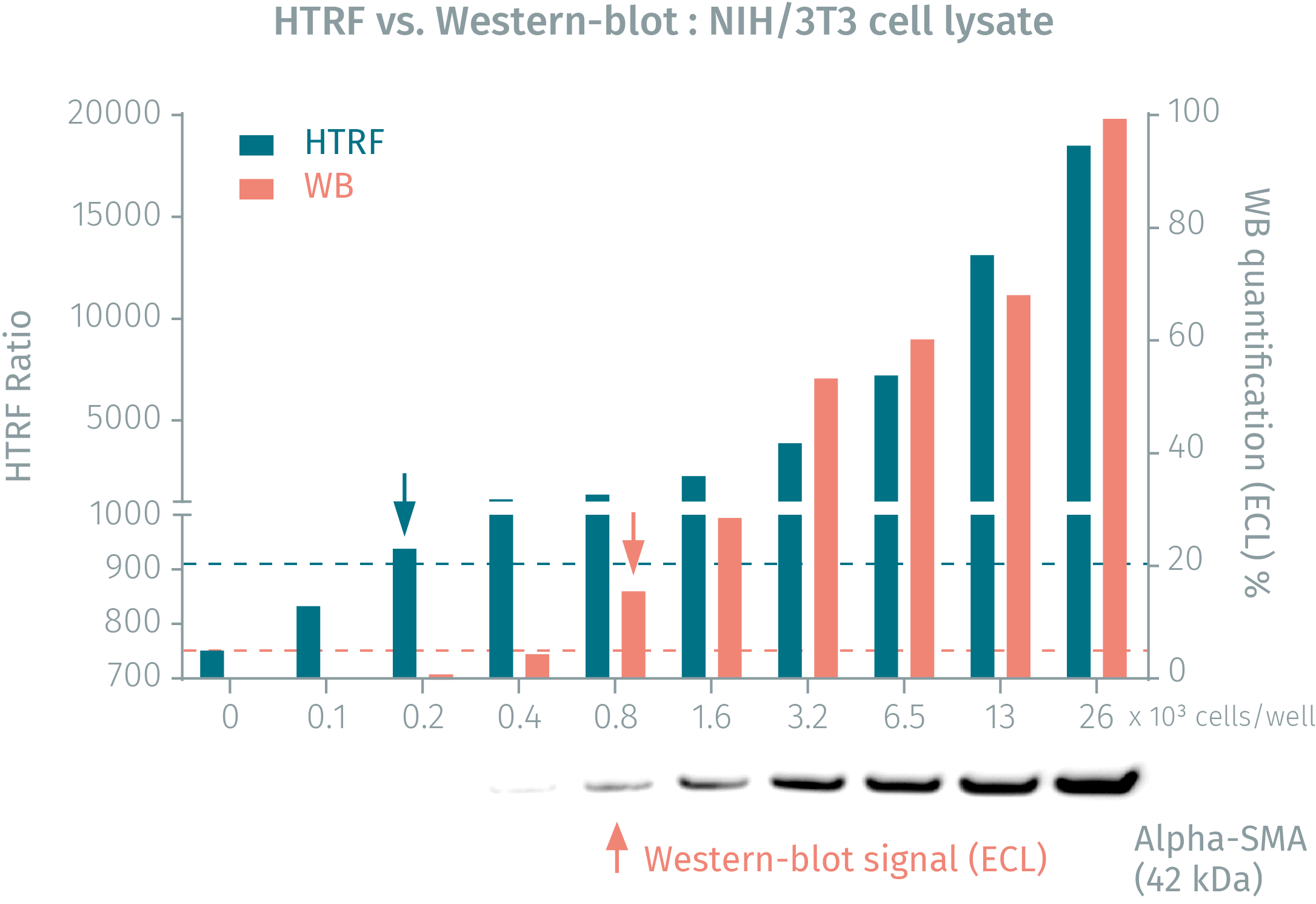
HTRF Alpha-SMA assay compared to Western Blot
NIH/3T3 cells were seeded in a T175 flask, and incubated for 2 days at 37°C. After medium removal, the cells were lysed for 30 minutes at RT under gentle shaking. Soluble supernatants were collected after a 10-minute centrifugation. Serial dilutions of the cell lysate were performed in the supplemented lysis buffer, and 16 µL of each dilution were transferred into a low volume white microplate before the addition of 4 µL of the HTRF® Alpha-SMA detection reagents. Equal amounts of lysates were used for a side-by-side comparison with Western Blot. Using HTRF® Alpha-SMA detection reagents, just 200 cells were sufficient for minimal signal detection, while 800 cells were needed to detect a significant Western Blot signal. Thus the HTRF® Alpha-SMA assay is 4-fold more sensitive than the Western Blot.
Simplified pathway
TGF-beta signaling pathway
TGF-beta is a profibrotic cytokine that represents the main myofibroblast inducer. The SMAD-dependent TGF-beta signaling pathway involving the activation of the transcription factors SMAD2/3 leads to ACTA2 gene transcription and expression of alpha-SMA (a-Smooth Muscle Actin). This specific actin isoform is incorporated into actin stress fibers and gives the cells a strong contractile activity characteristic of differentiated myofibroblasts. TGF-beta also signals through SMAD-independent pathways by activating the TAK1-dependent kinases p38, JNK and NF-kappaB, as well as AKT and ERK kinases.

Resources
Are you looking for resources, click on the resource type to explore further.
Guide
HTRF solutions, guide to major applications
This guide provides you an overview of HTRF applications in several therapeutic areas.
Guide
Improving immunotherapy by targeting the tumor stroma
Guide to How Cellular Actors of the Tumor Microenvironment Challenge Immunotherapies
Since the early 2000s, significant scientific...


How can we help you?
We are here to answer your questions.

