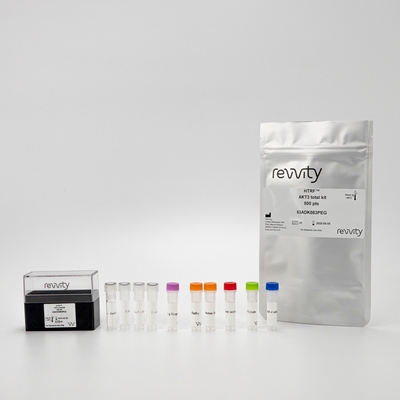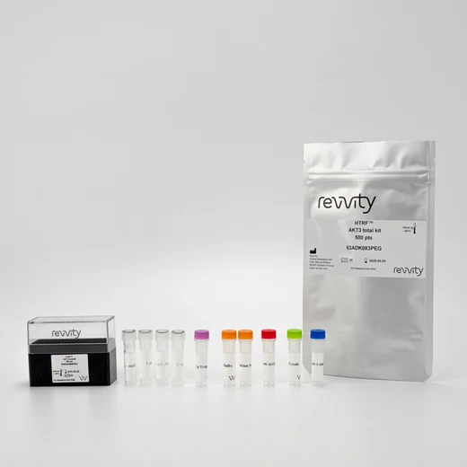

HTRF Human Total AKT3 Detection Kit, 10,000 Assay Points


HTRF Human Total AKT3 Detection Kit, 10,000 Assay Points


This HTRF kit is designed to monitor the expression level of cellular AKT3 and can be used as a normalization assay for the phospho AKT3 kit.
For research use only. Not for use in diagnostic procedures. All products to be used in accordance with applicable laws and regulations including without limitation, consumption and disposal requirements under European REACH regulations (EC 1907/2006).
| Feature | Specification |
|---|---|
| Application | Cell Signaling |
| Sample Volume | 16 µL |
This HTRF kit is designed to monitor the expression level of cellular AKT3 and can be used as a normalization assay for the phospho AKT3 kit.
For research use only. Not for use in diagnostic procedures. All products to be used in accordance with applicable laws and regulations including without limitation, consumption and disposal requirements under European REACH regulations (EC 1907/2006).


HTRF Human Total AKT3 Detection Kit, 10,000 Assay Points


HTRF Human Total AKT3 Detection Kit, 10,000 Assay Points


Product information
Overview
The Total AKT3 kit is designed to monitor the expression level of AKT3 and is used as a normalization assay with our phospho-AKT3 (Ser473) kits. Also known as protein kinase B (PKB), AKT is an oncogene playing a key role in controlling apoptosis, cell proliferation, transcription, cell migration, and glucose metabolism. AKT3 in particular plays a critical role in neuroscience.
Specifications
| Application |
Cell Signaling
|
|---|---|
| Brand |
HTRF
|
| Detection Modality |
HTRF
|
| Lysis Buffer Compatibility |
Lysis Buffer 1
|
| Molecular Modification |
Total
|
| Product Group |
Kit
|
| Sample Volume |
16 µL
|
| Shipping Conditions |
Shipped in Dry Ice
|
| Target Class |
Phosphoproteins
|
| Target Species |
Human
|
| Technology |
TR-FRET
|
| Therapeutic Area |
Neuroscience
|
| Unit Size |
10,000 Assay Points
|
Video gallery

HTRF Human Total AKT3 Detection Kit, 10,000 Assay Points

HTRF Human Total AKT3 Detection Kit, 10,000 Assay Points

How it works
Total-AKT3 assay principle
The Total-AKT3 assay quantifies the expression level of AKT3 in a cell lysate. Contrary to Western Blot, the assay is entirely plate-based and does not require gels, electrophoresis or transfer. The Total-AKT3 assay uses two labeled antibodies: one coupled to a donor fluorophore, the other to an acceptor. Both antibodies are highly specific for a distinct epitope on the protein. In presence of AKT3 in a cell extract, the addition of these conjugates brings the donor fluorophore into close proximity with the acceptor and thereby generates a FRET signal. Its intensity is directly proportional to the concentration of the protein present in the sample, and provides a means of assessing the proteins expression under a no-wash assay format.

Total-AKT3 2-plate assay protocol
The 2 plate protocol involves culturing cells in a 96-well plate before lysis then transferring lysates to a 384-well low volume detection plate before adding Total AKT3 HTRF detection reagents. This protocol enables the cells' viability and confluence to be monitored.

Total-AKT3 1-plate assay protocol
Detection of total AKT3 with HTRF reagents can be performed in a single plate used for culturing, stimulation and lysis. No washing steps are required. This HTS designed protocol enables miniaturization while maintaining robust HTRF quality.

Assay validation
Total AKT3 assay: inhibitory effect of blocking peptide
SH-SY5Y cells were stimulated with human IGF-1 for 15 min. After stimulation, media was removed and cells were lysed with lysis buffer 1X for 30 min at RT under gentle shaking. 14 µL of lysates were transferred into 384-well sv white microplates and 2 µL of different concentration of blocking peptide specific for AKT3 was added before adding 4 µL of the HTRF total AKT3 detection reagents. The HTRF signal was recorded after an overnight incubation at room temperature.

Simplified pathway
AKT simplified pathway
AKT (or protein kinase B) plays a key role in controlling survival and apoptosis. This serine/threonine protein kinase is regulated by insulin and various growth and survival factors, to work in a wortmannin-sensitive pathway involving PI 3 kinase. When the Pleckstrin Homology (PH) domain of AKT binds to phosphoinositides, AKT can be phosphorylated by two different kinases, PDK1 at threonine 308 and mTORC2 (mammalian target of rapamycin) at serine 473, which switch on AKT activation.

Resources
Are you looking for resources, click on the resource type to explore further.
Download this summary of signaling pathways in the immune system with HTRF targets.
Included:
- Detailed pathways about NK cells, T...


How can we help you?
We are here to answer your questions.


