


US
Revvity Sites Globally
Select your location.
*e-commerce not available for this region.

HTRF Human & Mouse Phospho-ERK (Thr202/Tyr204) Advanced Detection Kit, 500 Assay Points
")

HTRF Human & Mouse Phospho-ERK (Thr202/Tyr204) Advanced Detection Kit, 500 Assay Points
")

HTRF Human and Mouse Advanced phospho-ERK (Thr202/Tyr204) Detection Kit
")

This HTRF kit is designed for robust quantification of ERK modulation, phosphorylated at Thr202/Tyr204.
For research use only. Not for use in diagnostic procedures. All products to be used in accordance with applicable laws and regulations including without limitation, consumption and disposal requirements under European REACH regulations (EC 1907/2006).
| Feature | Specification |
|---|---|
| Application | Cell Signaling |
| Sample Volume | 16 µL |
This HTRF kit is designed for robust quantification of ERK modulation, phosphorylated at Thr202/Tyr204.
For research use only. Not for use in diagnostic procedures. All products to be used in accordance with applicable laws and regulations including without limitation, consumption and disposal requirements under European REACH regulations (EC 1907/2006).
Product Variants
Unit Size: 96 Assay Points
Part #:
64AERPET
List Price
USD 703.20
Unit Size: 500 Assay Points
Part #:
64AERPEG
List Price
USD 2,271.53
Unit Size: 10,000 Assay Points
Part #:
64AERPEH
List Price
USD 13,214.42
HTRF Human & Mouse Phospho-ERK (Thr202/Tyr204) Advanced Detection Kit, 500 Assay Points
HTRF Human and Mouse Advanced phospho-ERK (Thr202/Tyr204) Detection Kit
HTRF Human & Mouse Phospho-ERK (Thr202/Tyr204) Advanced Detection Kit, 500 Assay Points
Product information
Overview
The Advanced Phospho-ERK assay kits are designed for the robust and highly sensitive quantification of ERK phosphorylation on Thr202/Tyr204 as an MAPK pathway readout. The simple mix-and-read protocol eliminates all wash steps for faster analysis and high quality output. The Total-ERK1/2 assay is compatible with the buffers from the Phospho or Advanced phospho-ERK kits, so the same lysate can be used for analysis of the total and the phosphorylated protein populations. This kit is optimal for GPCR and RTK targeted screening.
Specifications
| Application |
Cell Signaling
|
|---|---|
| Brand |
HTRF
|
| Detection Modality |
HTRF
|
| Molecular Modification |
Phosphorylation
|
| Product Group |
Kit
|
| Sample Volume |
16 µL
|
| Shipping Conditions |
Shipped in Dry Ice
|
| Target Class |
Phosphoproteins
|
| Target Species |
Human
Mouse
|
| Technology |
TR-FRET
|
| Therapeutic Area |
Metabolism/Diabetes
NASH/Fibrosis
Neuroscience
Oncology & Inflammation
Rare Diseases
|
| Unit Size |
500 Assay Points
|
Video gallery
HTRF Human & Mouse Phospho-ERK (Thr202/Tyr204) Advanced Detection Kit, 500 Assay Points
HTRF Human & Mouse Phospho-ERK (Thr202/Tyr204) Advanced Detection Kit, 500 Assay Points
Citations
How it works
Advanced Phospho-ERK (Thr202/Tyr204) assay principle
The Phospho-ERK (Thr202/Tyr204) assay measures ERK when phosphorylated at Thr202/Tyr204. Contrary to Western Blot, the assay is entirely plate-based and does not require gels, electrophoresis or transfer. The Phospho-ERK (Thr202/Tyr204) assay uses 2 labeled antibodies: one with a donor fluorophore, the other one with an acceptor. The first antibody is selected for its specific binding to the phosphorylated motif on the protein, the second for its ability to recognize the protein independent of its phosphorylation state. Protein phosphorylation enables an immune-complex formation involving both labeled antibodies and which brings the donor fluorophore into close proximity to the acceptor, thereby generating a FRET signal. Its intensity is directly proportional to the concentration of phosphorylated protein present in the sample, and provides a means of assessing the proteins phosphorylation state under a no-wash assay format.
Advanced Phospho-ERK (Thr202/Tyr204) 2-plate assay protocol
The 2 plate protocol involves culturing cells in a 96-well plate before lysis then transferring lysates to a 384-well low volume detection plate before adding Phospho-ERK (Thr202/Tyr204) HTRF detection reagents. This protocol enables the cells' viability and confluence to be monitored.

Advanced Phospho-ERK (Thr202/Tyr204) 1-plate assay protocol
Detection of Phosphorylated ERK (Thr202/Tyr204) with HTRF reagents can be performed in a single plate used for culturing, stimulation and lysis. No washing steps are required. This HTS designed protocol enables miniaturization while maintaining robust HTRF quality.

Assay validation
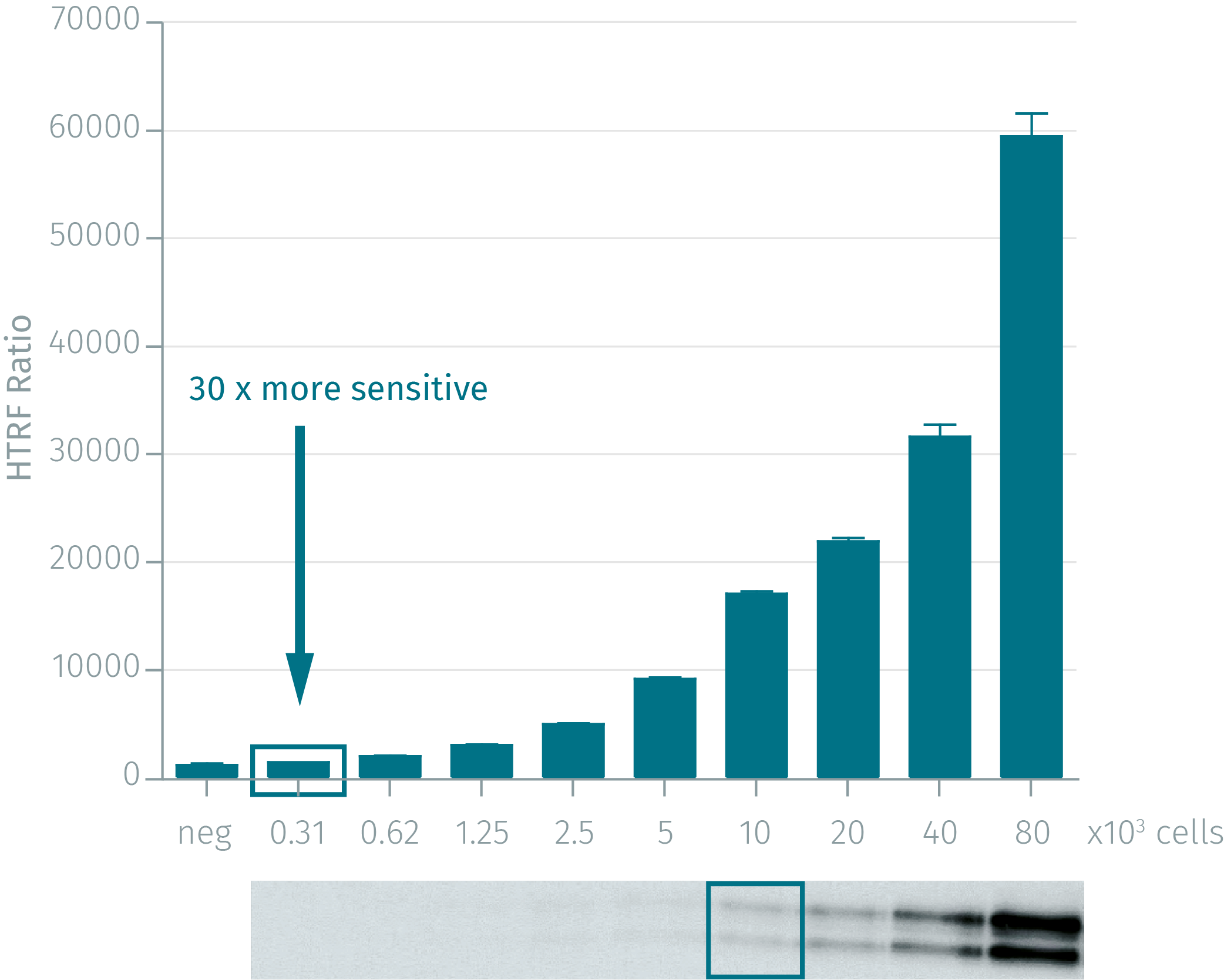
Unique sensitivity of Advanced phospho-ERK compared to WB
HEK293 cells were cultured for 48 h. Cells were then stimulated with 50 nM EGF for 5 min. After medium removal and lysis, cell lysates were collected and centrifuged for 10 minutes. Serial dilutions were performed and analyzed side-by-side by Western Blot and by HTRF Advanced phospho-ERK. By using HTRF Advanced phospho-ERK (Thr202/Tyr204) only 300 cells are sufficient for minimal signal detection, while 10,000 cells are needed for a Western Blot signal. The HTRF Advanced phospho-ERK assay is at least 30-fold more sensitive than the Western Blot, and shows optimal correlation.

Basal phospho-ERK dose response for inhibitors without stimulation
Advanced phospho-ERK enables the study of MAPK pathway inhibitor compounds on the basal level of activation, without the need of pathway stimulation. Due to its high cross reactivity, Advanced phospho-ERK is highly compatible with many different model species: human, mouse, rat, dog, monkey, hamster, mink, bovine, pig, D. Melanogaster and zebrafish. All experiments were performed using the two-plate assay protocol for adherent cells. One day after seeding, the cell lines were all treated with their corresponding inhibitor for 30 min at 37°C, 5% CO2.

Phospho-ERK modulation in diabetes models & patient blood cells
Advanced phospho-ERK assay kit is ideal for monitoring agonist stimulation in pancreatic beta-cell lines or patient PBMCs. The experiments were performed on two pancreatic beta-cell lines: Ins-1E and Min6. The two-plate assay protocol was utilized. Both cell lines were seeded at 70,000 cells/well for 4 days. Cells were washed and incubated with Krebs Ringer Buffer not containing glucose. After 2 hours of starvation, cells were stimulated with glucose. The PBMC's were dispensed at 100,000 cells/well and then stimulated with PMA for 30 min at 37°C, 5% CO2.

Sensitive phospho-ERK detection in tumor xenografts
BxPC-3 pancreatic tumor xenografts were excised from nude mice, ground and lysed. After centrifugation, the soluble fraction was collected and serial dilutions were performed prior to detection with HTRF Advanced phospho-ERK kit reagents.

Optimized cell density for phospho-ERK detection
Due to the high sensitivity of Advanced phospho-ERK, it is recommended to work with low cell densities of standard cell lines when expecting a high ERK phosphorylation level, to ensure operation in the linear range of the kit and to achieve an optimal S/B. Dose-response experiments were performed on HEK293 cells with the EGFR agonist EGF or the small molecule Raf kinase inhibitor L779-450, using the two-plate assay protocol for adherent cells. Cells were plated at 25,000, 50,000 or 100,000 cells/well for 24h at 37°C, 5% CO2.


Gaq/i coupled receptor activation
Results obtained on CHO-CCR5 (25 000 cells) activated with Rantes & MIP-Iß for 10', using the two-plate assay protocol of the phospho-ERK cellular assay

Screening robustness
CHO-M1 (5,000 cells/well - 384-well small volume plate) were stimulated with Carbachol for 10 minutes at RT. Z' values in fifty replicates were calculated between unstimulated cells (basal level) and 2 selected Carbachol concentrations, 1.3 µM and 4 µM, corresponding to the EC80 and EC100 respectively. Assay was performed using the One-plate assay protocol of the phospho-ERK kit on a robotic system from CyBio™
(CyBivario equipped with 384/25 µL pipeting head). Results were read on PHERAStar Plus (BMG Labtech).

Total-ERK1/2 assay to control phosphorylation of ERK1/2
A431 cells (100,000 cells/well) were activated with EGF for 10 min, using the two-plate assay protocol of the Phospho-ERK1/2 and Total-ERK1/2 assays. As expected, results obtained show a dose-response increase of ERK1/2 phosphorylation upon EGF stimulation while ERK1/2 expression level remains constant.

Simplified pathway
MAPK/ERK cell signaling
The MAPK/ERK signaling cascade is activated by a wide variety of receptors involved in growth and differentiation. The core signal transduction cascade elicits regulation of numerous cellular processes including adhesion, migration, apoptosis, differentiation and metabolism. MEK1/2 catalyze the phosphorylation of ERK1/2 at Tyr204 and Thr202. In response, ERK phosphorylates hundreds of cytoplasmic and nuclear substrates. The wide complexity and diversity of MAPK signaling makes ERK a key regulator and major signaling node in biology.

ERK in GPCR signaling
ERK modulation plays a major role in GPCR signaling and is therefore an important measurable GPCR readout. GPCRs act via G proteins to regulate a wide range of cellular functions. Upon stimulation, these receptors activate effectors like adenylate cyclase and phospholipase C, which influence not only intracellular concentrations of second messengers like cAMP and Ca2+, but also mediate ERK1/2 phosphorylation. GPCRs have also been shown to mediate ERK1/2 activation in a G protein-independent but beta-arresting dependent manner.
Resources
Are you looking for resources, click on the resource type to explore further.
Guide
HTRF solutions, guide to major applications
This guide provides you an overview of HTRF applications in several therapeutic areas.
Guide
Understanding obesity: exploring cellular pathways and mechanisms
Obesity is a complex condition characterized by excessive fat accumulation, posing significant health and socioeconomic challenges...


How can we help you?
We are here to answer your questions.

