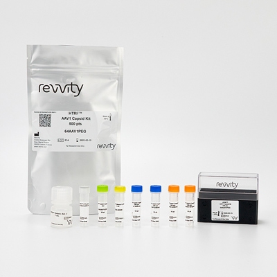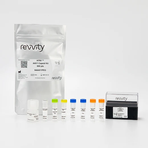

HTRF AAV1 Detection Kit, 500 Assay Points


HTRF AAV1 Detection Kit, 500 Assay Points


The AAV1 Capsid kit is designed for the quantitative measurement of Adeno-associated virus serotype 1 (AAV1) particles in both cell lysates and cell supernatants.
For research use only. Not for use in diagnostic procedures. All products to be used in accordance with applicable laws and regulations including without limitation, consumption and disposal requirements under European REACH regulations (EC 1907/2006).
| Feature | Specification |
|---|---|
| Application | Bioprocessing |
| Sample Volume | 5 µL |
The AAV1 Capsid kit is designed for the quantitative measurement of Adeno-associated virus serotype 1 (AAV1) particles in both cell lysates and cell supernatants.
For research use only. Not for use in diagnostic procedures. All products to be used in accordance with applicable laws and regulations including without limitation, consumption and disposal requirements under European REACH regulations (EC 1907/2006).


HTRF AAV1 Detection Kit, 500 Assay Points


HTRF AAV1 Detection Kit, 500 Assay Points


Product information
Overview
Adeno-associated virus (AA) vectors are the leading platform for gene delivery for the treatment of a variety of human diseases. AAV Serotype 1 (AAV1) is an efficient vector for gene delivery to skeletal muscle or the central nervous system. The AAV1 kit is designed to detect and quantify AAV1 particles in an easy-to-use, no-wash format. The simple and robust procedure benefits from increased throughput compared to ELISA.
Specifications
| Application |
Bioprocessing
|
|---|---|
| Brand |
HTRF
|
| Detection Modality |
HTRF
|
| Lysis Buffer Compatibility |
Lysis Buffer 1
Lysis Buffer 2
Lysis Buffer 3
Lysis Buffer 4
|
| Product Group |
Kit
|
| Sample Volume |
5 µL
|
| Shipping Conditions |
Shipped in Dry Ice
|
| Target Class |
Viral Particles
|
| Technology |
TR-FRET
|
| Unit Size |
500 Assay Points
|
Video gallery

HTRF AAV1 Detection Kit, 500 Assay Points

HTRF AAV1 Detection Kit, 500 Assay Points

How it works
AAV1 Capsid assay principle
The Adeno-Associated Virus serotype 1 (AAV1) assay measures AAV1 capsid in cell supernatant or cell lysate. The assay uses two anti-AAV1 antibodies: one coupled to HRP and which binds to anti-HRP d2 (acceptor) in premix 1, and the other coupled to biotin and which binds to Streptavidin Eu-cryptate (donor) in premix 2. In presence of AAV1 capsid in a cell extract or supernatant, the addition of these conjugates brings the donor fluorophore into close proximity with the acceptor, and thereby generates a FRET signal. Its intensity is directly proportional to the concentration of the capsid present in the sample, and provides a means of assessing any changes caused by experimental variability under a no-wash assay format.

AAV1 Capsid assay protocol
The AAV1 Capsid assay protocol using a 384-well small volume white plate is described on the right. 5 µL of sample or standard and 5 µL of diluent are dispensed directly into the plate for detection by HTRF® reagents. The Biotin antibody anti-AAV1 is pre-mixed with Streptavidin labeled with the donor, and HRP anti-AAV1 was pre-mixed with anti-HRP labeled with the acceptor. 5 µL of each premix were added. The assay can be run in up to a 1536-well format by simply resizing each addition volume proportionally.

Assay details
standard curve Spike S1 kit
| Sample size | 5 µL |
|---|---|
| Final assay volume | 20 µL |
| Time to result | Overnight at RT |
| Detection limit (LOD) in diluent | 2.76E+08 VP/mL |
| Dynamic range | 9.73E+08 – 2.50E+11 VP/mL |
| Sample compatibility |
From raw harvest material to the final product Supernatant, Cell Lysate (LB#3) |

Analytical performance
Precision
Intra assay (n=24)
| Sample | Mean [AAV1] (VP/mL) | CV |
|---|---|---|
| 1 | 2.00E+11 | 4% |
| 2 | 5.00E+10 | 5% |
| 3 | 1.25E+10 | 4% |
| Mean CV | 4% |
Each of the 3 samples was measured 24 times, and % CV was calculated for each sample.
Inter assay (n=4)
| Sample | Mean [AAV1] (VP/mL) | CV |
|---|---|---|
| 1 | 2.00E+11 | 2% |
| 2 | 5.00E+10 | 4% |
| 3 | 1.25E+10 | 8% |
| Mean CV | 4% |
Each of the samples was measured in 3 independent experiments performed by different operators, and CV percentages were calculated for each sample.
Dilutional linearity
|
Dilution Factor |
[AAV1] Expected (VP/mL) |
[AAV1] Mesured (VP/mL) |
Dilution Recovery |
|---|---|---|---|
|
Neat |
- |
1.61E+11 |
100% |
|
2 |
8.03E+10 |
7.38E+10 |
109% |
|
4 |
4.01E+10 |
3.46E+10 |
116% |
|
8 |
2.01E+10 |
1.73E+10 |
116% |
|
16 |
1.00E+10 |
9.26E+09 |
108% |
|
Mean |
|
|
110% |
Interferences
|
[AAV1] (VP/mL) |
[Total Protein] SF9 cell lysates Spiked Sample (mg/mL) |
Recovery |
|---|---|---|
|
1.0E+11 |
0.5 |
54% |
|
0.375 |
59% |
|
|
0.25 |
70% |
|
|
0.125 |
81% |
|
|
0.05 |
93% |
|
[AAV1] (VP/mL) |
[Total Protein] HEK293 cell lysates Spiked Sample (mg/mL) |
Recovery |
|---|---|---|
|
1.0E+11 |
1.5 |
64% |
|
0.75 |
96% |
|
|
0.5 |
96% |
|
|
0.375 |
94% |
|
|
0.25 |
103% |
Antigen Spike and Recovery
|
Sample |
[AAV1] Standard (VP/mL) |
SF9 cell lysate (0.05 mg/ml) |
|---|---|---|
|
Recovery |
||
|
1 |
1.00E+11 |
93% |
|
2 |
5.00E+10 |
96% |
|
3 |
1.00E+10 |
101% |
|
|
Mean CV |
97% |
|
Sample |
[AAV1] Standard (VP/mL) |
HEK293 cell lysate (0.75 mg/ml) |
|---|---|---|
|
Recovery |
||
|
1 |
1.00E+11 |
96% |
|
2 |
5.00E+10 |
98% |
|
3 |
5.00E+09 |
93% |
|
|
Mean CV |
96% |
Cross reactivities
Cross reactivities were assessed using other serotypes from the AAVs family. Standard curves were generated for each serotype using AAVs capsids diluted in the kit diluent. 5 µL of capsids were transferred into a white detection plate (384 low volume), and 5 µL of diluent followed by 10 µL of the HTRF AAV1 capsid detection reagents were added. The HTRF signal was recorded after an overnight incubation at room temperature. The assay showed differential affinities depending on the serotype, but did not detect AAV8 and AAV9.

Assay validation
Validation of HTRF AAV1 capsid detection on full and empty AAV1 particles
To demonstrate the detection of both full and empty AAV1 capsids, recognition of full AAV1-CMV-eGFP and empty AAV1 capsids were analyzed in the assay. A large range of AAV1-CMV-eGFP concentrations (GC/mL) were converted into VP/mL using an independent sample quantitation assay. 5 µL of full or empty capsids diluted in the kit diluent were then transferred into a white detection plate (384 low volume), and 5 µL of diluent followed by 10 µL of the HTRF AAV1 capsid detection reagents were added. The HTRF signal was recorded after an overnight incubation at room temperature. As expected, the HTRF AAV1 capsid detection assay could detect both full and empty AAV1 capsids in the same way.

Resources
Are you looking for resources, click on the resource type to explore further.
When a genetic mutation occurs, it can lead to the development of different pathologies. Over the past 10 years exciting...


How can we help you?
We are here to answer your questions.


