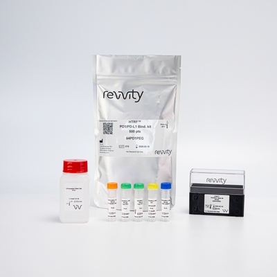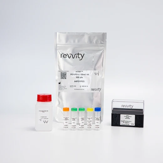

HTRF Human PD1 / PDL1 Binding Kit, 500 Assay Points


HTRF Human PD1 / PDL1 Binding Kit, 500 Assay Points


This immune checkpoint assay is designed to identify human PD1/PD-L1 checkpoint inhibitors.
| Feature | Specification |
|---|---|
| Application | Protein-Protein Interaction |
| Sample Volume | 2 µL |
This immune checkpoint assay is designed to identify human PD1/PD-L1 checkpoint inhibitors.


HTRF Human PD1 / PDL1 Binding Kit, 500 Assay Points


HTRF Human PD1 / PDL1 Binding Kit, 500 Assay Points


Product information
Overview
Programmed cell death protein 1 (PD1) is an immune checkpoint receptor that regulates T cell response. Its ligand, programmed death-ligand 1 (PD-L1), is commonly over-expressed on a tumor cell surface. When PD1 is bound to PD-L1, T cell response is suppressed, contributing to tumor immune resistance. Checkpoint inhibitors blocking PD1/PD-L1 complex formation are generating considerable interest in cancer immunotherapy.
Specifications
| Application |
Protein-Protein Interaction
|
|---|---|
| Brand |
HTRF
|
| Detection Modality |
HTRF
|
| Product Group |
Kit
|
| Sample Volume |
2 µL
|
| Shipping Conditions |
Shipped in Dry Ice
|
| Target Class |
Binding Assay
|
| Target Species |
Human
|
| Technology |
TR-FRET
|
| Therapeutic Area |
Oncology & Inflammation
|
| Unit Size |
500 Assay Points
|
Video gallery

HTRF Human PD1 / PDL1 Binding Kit, 500 Assay Points

HTRF Human PD1 / PDL1 Binding Kit, 500 Assay Points

Citations
How it works
Assay principle
The PD1/PD-L1 binding assay includes tagged human recombinant immune checkpoint partners (PD1 and PD-L1) and labelled anti-tag reagents for HTRF detection. Without an inhibitor, PD1 binds to PD-L1, and the binding of each detection reagent to its tagged target generates an HTRF signal. In the presence of compound, standard, or an antibody blocking the PD1 /PD-L1 interaction, the HTRF signal decreases.

Assay protocol
This PD1/PD-L1 binding assay can be run in a 96- or 384-well low volume white plate (20 µL final). As described here, samples or standards are dispensed directly into the assay plate, and the tagged PD1 & PD-L1 proteins are then added, followed by the dispensing of the HTRF reagents. The reagents labelled with HTRF fluorophores may be pre-mixed and added in a single dispensing step. No washing steps are needed. The protocol can be further miniaturized or upscaled by simply resizing each addition volume proportionally.

Assay validation
Inhibitory effect of blocking antibodies and small molecules
The inhibitory effects of three antibodies and two small molecules known to inhibit the PD1 / PD-L1 interaction were tested. The two anti-PD1 antibodies (pembrolizumab & nivolumab) were shown to inhibit the interaction with IC50 values of 2.0 & 2.4 nM respectively. The anti-PD-L1 antibody (atezolizumab) disrupted the interaction, with an IC50 of 3.9 nM. Two small molecule PD1 / PD-L1 inhibitors 1 & 3 show IC50s of 177 & 146 nM in these assay conditions.

Inhibitory effect of the PD1 / PD-L1 standard
The PD1 / PD-L1 standard stock solution provided in the kit was diluted to prepare the 8-point dose response curve using 5-fold serial dilutions. As described in the assay protocol, standards were dispensed into the assay plate, and the tagged PD1 & PD-L1 proteins were then added, followed by the dispensing of the HTRF reagents. The data presented in the graph show that the PD1 / PD-L1 interaction is inhibited, with an IC50 of 3.8 nM.

Resources
Are you looking for resources, click on the resource type to explore further.
A single White Paper for a review of immuno-oncology
Immuno-oncology, the field which stimulates a individual's own immunity in...
Discover the versatility and precision of Homogeneous Time-Resolved Fluorescence (HTRF) technology. Our HTRF portfolio offers a...
This guide provides you an overview of HTRF applications in several therapeutic areas.
A healthy immune system protects the body, but dysfunction can lead to autoimmune diseases affecting various organs. Despite over...
Advance your autoimmune disease research and benefit from Revvity broad offering of reagent technologies
Chimeric antigen receptor (CAR) T-cell therapy has transformed the field of immuno-oncology providing a novel approach to treating...
Loading...


How can we help you?
We are here to answer your questions.


