Menu
NL
NL

G protein-coupled receptors (GPCRs) are the largest family of membrane proteins and the most studied targets in drug discovery. They relay extracellular signals by triggering intracellular pathways through coupling with G proteins and arrestins.

Revvity offers a wide panel of GPCR signaling assays to support your research. 

Our large range of GPCR assays allows you to study every step of signal transduction, including:

  • Ligand binding (Tag-lite® and radioactive ligand)
  • G-protein activation (Gs, Gq, Gi) through cAMP, GTP and IP-one
  • Arrestins recruitment
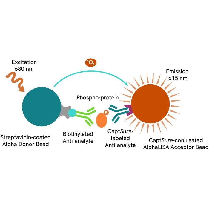
  • Intracellular signaling with phospho-assays (AKT, ERK, CREB, MEK…).

Discover our GPCR research reagents portfolio with solutions spanning a huge variety of research applications.

Introduction-gpcr-overview-image1_800.png
What you need to know about GPCRs

GPCRs are cell membrane proteins that consist of seven membrane-spanning domains. They are activated by external signals coming from ligands (hormones, neurotransmitters, ions…) and are the main broker in the transmission of those external signals to the inside of cells.

GPCRs are the largest family of targeted proteins and represent more than 34% of all FDA-approved therapeutic drug targets. They play a critical role in many physiological and cellular processes such as immunity, metabolism, and cell development and proliferation, and are involved in numerous therapeutic areas.

Introduction-gpcr-therapeutic-area-image2_800.png

Despite GPCRs being a well-known drug target class, they continue to offer scope for the development of new therapeutics. Many GPCRs remain undescribed, both in function and interaction with ligands. These undescribed receptors are called “orphans” and their investigation holds great promise for the future of therapeutic research.

GPCR Research Reagents
1283450_GPCR-category-page-rvty-1

Ligand binding

The ligand/receptor binding is the first key step in GPCR signaling. Revvity offers a range of ligand/receptor interaction solutions in both radioactive format and fluorescence assays with our proprietary HTRF-based Tag-lite technology.

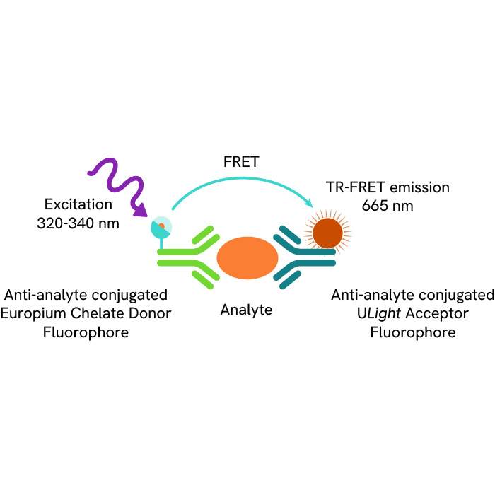
  • Tag-lite - Tag-lite is a non-radioactive TR-FRET-based solution which can be used to assess the pharmacology and pharmacodynamics of ligand/receptor interactions. The receptor of interest is labeled with cryptate in a way that does not alter receptor binding. The corresponding ligand is labeled with d2 acceptor. When the d2-labeled ligand binds to the cryptate-labeled receptor, it produces a HTRF signal.

    In a competition assay, the introduction of a competing small molecule dislodges the d2-labeled ligand. Consequently, the ligand is pushed away and the signal stops.

    ligand-binding-image1_800.png


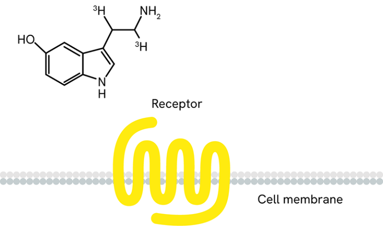
  • Receptor ligand binding with radiolabeled ligands and GPCR membranes - We also offer a range of radioactive ligands, which are used to study molecular interactions and QC our GPCR over-expressing cell lines and membrane preparations. Radiometric ligand binding assays are conducted on cells or cell membranes containing a GPCR receptor of interest. Radioligands can be used to perform saturation curves, competition, and kinetic experiments.
    ligand-binding-image2_800.png


  • Radioactive ligand binding assays can be performed in several different formats. Typically, we perform this assay in filtration format, where the unbound ligand is washed away using a vacuum manifold or cell harvester. The assay can also be conducted in a homogenous Scintillation Proximity Assay (SPA) format, where no wash steps are required.

    Over 100 GPCR-expressing membrane preparations are validated for receptor ligand binding and over 50 are validated for GTPγS binding.

The ligand/receptor binding is the first key step in GPCR signaling. Revvity offers a range of ligand/receptor interaction solutions in both radioactive format and fluorescence assays with our proprietary HTRF-based Tag-lite technology.

  • Tag-lite - Tag-lite is a non-radioactive TR-FRET-based solution which can be used to assess the pharmacology and pharmacodynamics of ligand/receptor interactions. The receptor of interest is labeled with cryptate in a way that does not alter receptor binding. The corresponding ligand is labeled with d2 acceptor. When the d2-labeled ligand binds to the cryptate-labeled receptor, it produces a HTRF signal.

    In a competition assay, the introduction of a competing small molecule dislodges the d2-labeled ligand. Consequently, the ligand is pushed away and the signal stops.

    ligand-binding-image1_800.png


  • Receptor ligand binding with radiolabeled ligands and GPCR membranes - We also offer a range of radioactive ligands, which are used to study molecular interactions and QC our GPCR over-expressing cell lines and membrane preparations. Radiometric ligand binding assays are conducted on cells or cell membranes containing a GPCR receptor of interest. Radioligands can be used to perform saturation curves, competition, and kinetic experiments.

    ligand-binding-image2_800.png


  • Radioactive ligand binding assays can be performed in several different formats. Typically, we perform this assay in filtration format, where the unbound ligand is washed away using a vacuum manifold or cell harvester. The assay can also be conducted in a homogenous Scintillation Proximity Assay (SPA) format, where no wash steps are required.

    Over 100 GPCR-expressing membrane preparations are validated for receptor ligand binding and over 50 are validated for GTPγS binding.
1283450_GPCR-category-page-rvty-2

G-protein activation

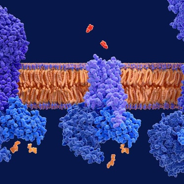
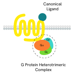
The main signal transduction of GPCRs is dependent on the receptor-mediated activation of heterotrimeric G-proteins.

G-proteins are composed of three subunits (Gα, Gβ and Gγ) and are classified into four families (Gs, Gi/o, Gq/11, and G12/13):

  • Gαs-coupled GPCRs positively stimulate the activity of adenylate cyclase, resulting in an increase in cellular cAMP
  • Gαi-coupled GPCRs lead to a negative regulation of adenylate cyclase, and thus to a decrease in cAMP production
  • Gαq protein signaling is based on enzymes of the phospholipase C family (PLC).

    gprotein-activation-image1_800.png

The main signal transduction of GPCRs is dependent on the receptor-mediated activation of heterotrimeric G-proteins.

G-proteins are composed of three subunits (Gα, Gβ and Gγ) and are classified into four families (Gs, Gi/o, Gq/11, and G12/13):

  • Gαs-coupled GPCRs positively stimulate the activity of adenylate cyclase, resulting in an increase in cellular cAMP
  • Gαi-coupled GPCRs lead to a negative regulation of adenylate cyclase, and thus to a decrease in cAMP production
  • Gαq protein signaling is based on enzymes of the phospholipase C family (PLC).

    gprotein-activation-image1_800.png
1283450_GPCR-category-page-rvty-3

Arrestins recruitment

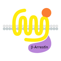
GPCRs also initiate G-protein-independent pathways that instead rely on arrestin coupling, which in turn suppresses G-protein activation.

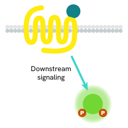
When a ligand binds to the extracellular part of a GPCR, the receptor undergoes conformational changes and opens its intracellular part to be phosphorylated by kinases. This phosphorylation of GPCRs by GRKs (GPCR kinases) is a prerequisite for high-affinity arrestin binding. The phosphorylated GPCR becomes a binding site for arrestins. Once linked, arrestins can interact with several partners such as AP2 or signaling proteins.

arrestin-recruitment-image1_800.png

GPCRs also initiate G-protein-independent pathways that instead rely on arrestin coupling, which in turn suppresses G-protein activation.

When a ligand binds to the extracellular part of a GPCR, the receptor undergoes conformational changes and opens its intracellular part to be phosphorylated by kinases. This phosphorylation of GPCRs by GRKs (GPCR kinases) is a prerequisite for high-affinity arrestin binding. The phosphorylated GPCR becomes a binding site for arrestins. Once linked, arrestins can interact with several partners such as AP2 or signaling proteins.

arrestin-recruitment-image1_800.png
1283450_GPCR-category-page-rvty-4

Intracellular signaling

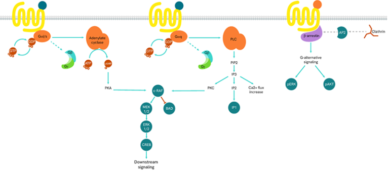
GPCRs are at the beginning of many phosphorylation cascades involving phosphatases bound to second messengers.

Following the activation of a GPCR, signals are sent via G-proteins and arrestins to second messengers. Each GPCR has its own signaling pathway, but kinases and phosphatases are always involved and promote phosphorylation cascades to the nucleus. These molecular events are implicated in many physiological and cellular processes such as cell survival, proliferation, differentiation, and metabolism.
Intracellular-signaling-image1_800.png

GPCRs are at the beginning of many phosphorylation cascades involving phosphatases bound to second messengers.

Following the activation of a GPCR, signals are sent via G-proteins and arrestins to second messengers. Each GPCR has its own signaling pathway, but kinases and phosphatases are always involved and promote phosphorylation cascades to the nucleus. These molecular events are implicated in many physiological and cellular processes such as cell survival, proliferation, differentiation, and metabolism.

Intracellular-signaling-image1_800.png
bg-element-listing-page bg-element-listing-page-mobile
Products & Services
Resource Library
Filters
Expand All|Collapse All
Product Type
Application
Brand
Cellular or Signaling Pathway
Concentration
Detection Modality
Host Species
Product Group
Radioisotope
Receptor
Target
Target Class
Target Species
Therapeutic Area
Validation
Volume
Sort by: Recommended
Recommended
Product Name A-Z
Product Name Z-A
Price - low to high
Price - high to low
1 - 25 of 237 Products and Services
Receptor cell membranes

Cell membranes prepared from HEK293 cells stably transfected with human opioid kappa (OP2) receptor, 400 units.

Part Number: 6110558400UA
EUR 1,182.00
Receptor cell membranes

Cell membranes prepared from CHO-K1 cells stably-transfected with human Nociceptin NOP (ORL1) receptor, 400 units.

Part Number: 6110540400UA
EUR 1,237.00
Receptor cell membranes

Cell membranes prepared from cells stably-transfected with human Vasopressin V1b receptor, in HEK-293 cells.

Part Number: RBHV1BM400UA
EUR 1,207.00
Receptor cell membranes

Cell membranes prepared from cells stably-transfected with human Cannabinoid CB2 receptor, in HEK-293 cells.

Part Number: RBXCB2M400UA
EUR 1,180.00
Shipping box for Revvity reagent kits

This HTRF kit is designed for robust cell-based quantification of AKT2 modulation, phosphorylated on Ser473.

Part Number: 63ADK080PEG, 63ADK080PEH
EUR 1,924.00 - 12,462.00
Receptor cell membranes

Cell membranes prepared from HT29 cells containing human endogenous Vasoactive Intestinal Polypeptide (VIP) receptor.

Part Number: RBHVIPM400UA
EUR 1,267.00
Receptor cell membranes

Cell membranes prepared from CHO-K1 cells stably-transfected with human serotonin 5HT7 receptor, 400 units.

Part Number: 6110512400UA
EUR 1,214.00
Shipping box for Revvity reagent kits

The Total Beta-arrestin 2 kit enables the cell-based quantitative detection of beta-arrestin 2, for monitoring GPCR activity.

Part Number: 64BAR2TPEB, 64BAR2TPEC
EUR 2,827.00 - 18,630.00
Receptor cell membranes

Cell membranes prepared from HEK293 cells stably transfected with human GABA B1a receptor, 400 units.

Part Number: 6110560400UA
EUR 1,238.00
Receptor cell membranes

Cell membranes prepared from HEK293 cells stably transfected with human GABA B1b receptor, 400 units.

Part Number: 6110557400UA
EUR 1,238.00
Receptor cell membranes

Cell membranes prepared from CHO-K1 cells stably-transfected with human Vasopressin V1b receptor, 400 units.

Part Number: 6110543400UA
EUR 1,206.00
Receptor cell membranes

Cell membranes prepared from cells stably-transfected with human Vasopressin V1a receptor, in HEK-293 cells.

Part Number: RBHV1AM400UA
EUR 1,210.00
Receptor cell membranes

Cell membranes prepared from CHO-K1 cells stably-transfected with human GABA B1b receptor, 400 units.

Part Number: 6110546400UA
EUR 1,204.00
Receptor cell membranes

Cell membranes prepared from cells stably-transfected with human serotonin 5HT1a receptor

Part Number: 6110501400UA
EUR 1,218.00
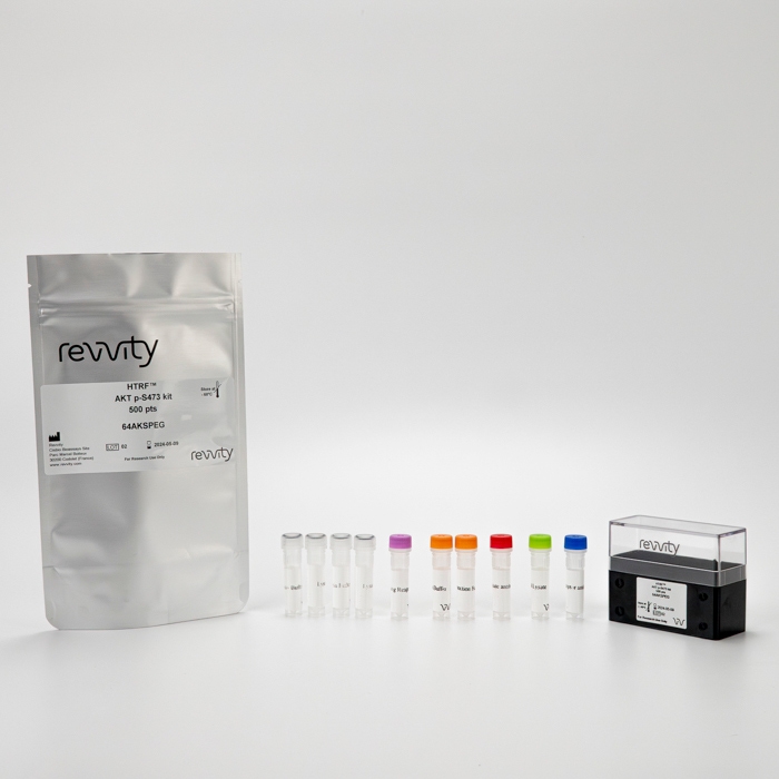
PHOTO AKT PHOSPHO-S473 KIT

This HTRF kit is designed to monitor pan Akt phosphorylation on Ser473 as a readout of Pi3K pathway activation.

Part Number: 64AKSPET, 64AKSPEG, 64AKSPEH
EUR 680.00 - 12,462.00
Shipping box for Revvity reagent kits

Mek1/2 P-s218/222 Kit - 50,000 Tests

Part Number: 64ME2PEG, 64ME2PEH
EUR 1,924.00 - 12,462.00
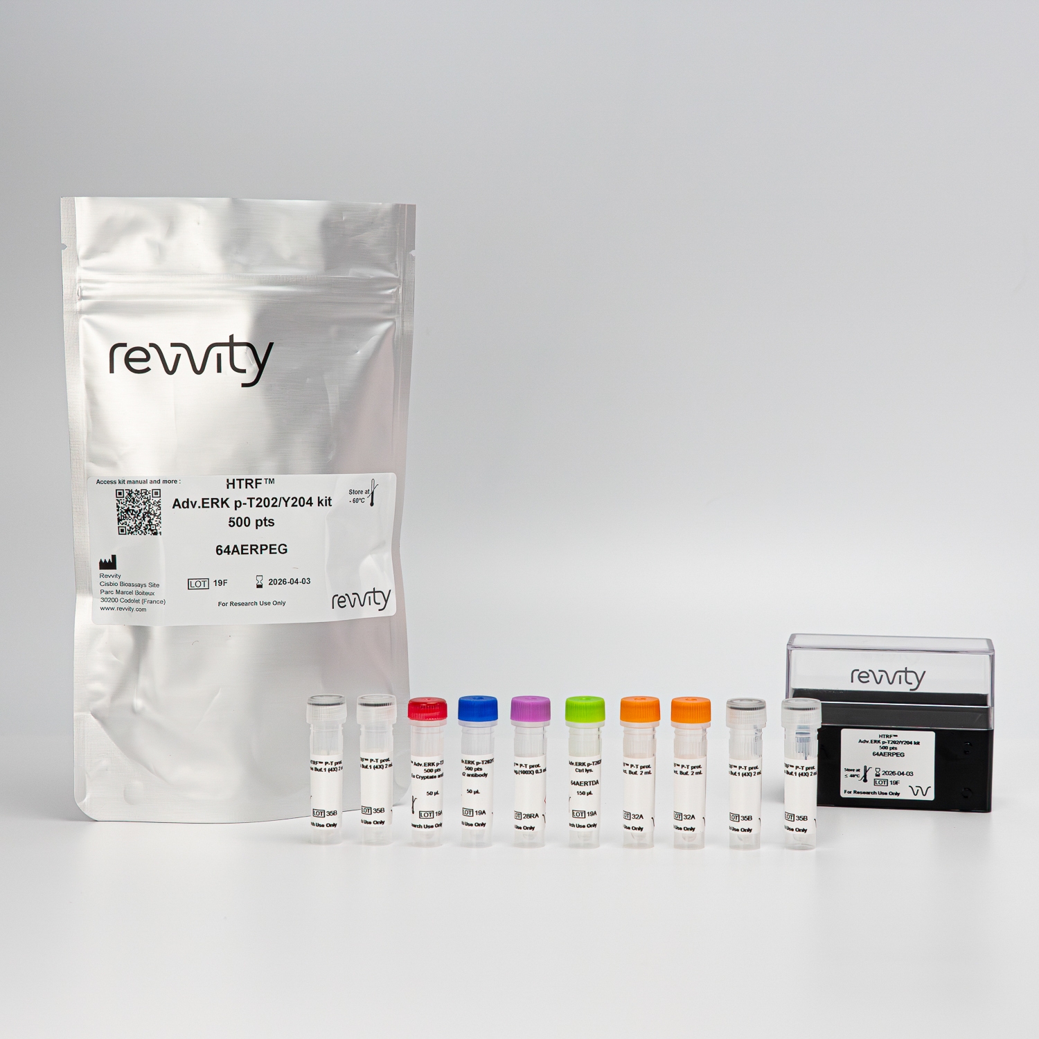
HTRF Human and Mouse Phospho-ERK (Thr202/Tyr204) Detection Kit

The phospho-ERK (Thr202/Tyr204) kit measures ERK protein phophorylated at Thr202/Tyr204 as a readout of MAPK pathway activation.

Part Number: 64ERKPEG, 64ERKPEH
EUR 1,924.00 - 12,462.00
PHOTO TOTAL AKT 1

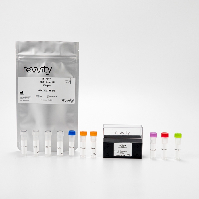
This HTRF kit is designed to monitor the expression level of cellular AKT1 and can be used as a normalization assay for the phospho-AKT1 kit.

Part Number: 63ADK079PEG, 63ADK079PEH
EUR 1,924.00 - 12,462.00
Receptor cell membranes

Our Membrane Target Systems: Melatonin MT1 (human) membranes are prepared from cells that express recombinant or endogenous receptors. Selected for optimal performance in binding assays.

Part Number: ES-620-M400UA
EUR 1,117.00
Lance Ultra Detection Kit

The LANCE® Ultra cAMP assay is a homogeneous time-resolved fluorescence resonance energy transfer (TR-FRET) immunoassay designed to measure cAMP produced upon modulation of adenylyl cyclase activity by G-protein coupled receptors (GPCRs).

Part Number: TRF0262, TRF0263, TRF0264
EUR 826.00 - 10,722.00
Receptor cell membranes

Wild Type Subtype A9L (mouse) Control Membrane 10mg Frozen suspension cell membrane. Shipped on dry ice.

Part Number: RBMA9M010MG
EUR 719.00
AlphaLISA SureFire Ultra Phospho-Protein image

The AlphaLISA™ SureFire® Ultra™ p-CREB (Ser133) assay is a sandwich immunoassay for quantitative detection of phospho-CREB (phosphorylated on Ser133) in cellular lysates using Alpha Technology.

EUR 746.00 - 47,567.00
Receptor cell membranes

Wild Type Subtype U-373 (human) Control Membrane Frozen suspension cell membrane. Shipped on dry ice.

Part Number: RBHU37M010MG
EUR 719.00
Receptor cell membranes

Our Membrane Target Systems: Melatonin MT2 (human) membranes are prepared from cells that express recombinant or endogenous receptors. Selected for optimal performance in binding assays.

Part Number: ES-621-M400UA
EUR 1,116.00
Receptor cell membranes

Cell membranes prepared from cells stably-transfected with human Melanocortin MC3 receptor, in HEK-293 cells.

Part Number: RBXMC3M400UA
EUR 1,205.00
Filters
Expand All|Collapse All
Resource Type
Sort by: Resource Name A-Z
Resource Name A-Z
Resource Name Z-A
1 - 25 of 50 Resources
Application Note
Application Note
A guideline for HTRF cell-based phospho-protein data normalization

Get the best out of your phosphorylation assays Combining phospho and total protein assays enables better analysis. This Application Note provides valuable guidelines for efficiently analyzing and interpreting results from such assay combinations. Check out all the tips and examples in features! Features Introduction to phospho and total protein assay relevance Tips for handling and interpreting data Examples from actual experiments

Technical Note
Technical Note
A simple method for preparing GPCR membrane model extracts from stable cell lines for use with the HTRF GTP Gi binding assay

G-protein coupled receptors (GPCRs) are crucial transmembrane proteins involved in cellular signal transduction. This technical note outlines a method for preparing GPCR membrane model extracts from stable cell lines, specifically for use with the HTRF GTP Gi binding assay. Get this technical note and discover: Key Highlights such as the Importance of GPCRs and the advantages of using HTRF GTP Gi Binding Assay Detailed Method with Cell Culture Preparation, Cell Lysis, Membrane Preparation and Assay Optimization For research use only. Not for use in diagnostic procedures.

Brochure
Brochure
Alpha SureFire Ultra no-wash immunoassay catalog

Discover Alpha SureFire ®   Ultra ™ assays, the no-wash cellular kinase assays leveraging Revvity's exclusive bead-based technology and sandwich immunoassays for detecting phosphorylated proteins in cells. Offering a quantitative alternative to Western Blotting, Alpha SureFire assays are automation-friendly, easily miniaturized, and proficient in detecting both endogenous and recombinant proteins. Explore our comprehensive portfolio of SureFire Assays, designed to help you elevate and expedite your drug discovery journey.

Guide
Guide
AlphaLISA SureFire Ultra assay optimization

This guide outlines further possible optimization of cellular and immunoassay parameters to ensure the best possible results are obtained.

Whitepaper
Whitepaper
An overview of atherosclerosis

Atherosclerosis pathogenesis, cellular actors, and pathways Atherosclerosis is a common condition in which arteries harden and become narrow due to a build-up of fatty material, usually cholesterol, and other substances such as calcium. This can lead to a range of serious health complications, including heart attack or stroke, making the disease an important contributing factor in death and morbidity in developed countries. Recent developments in our understanding of atherosclerosis from a molecular perspective include the discovery of new players in disease pathogenesis. Included in this white paper Atherosclerosis: step-by-step pathogenesis, therapeutic strategies, and recent developments Detailed descriptions and explanations, including a focus on pathways

Product Info
Product Info
Calendar: NEN® Radiochemicals Fresh Lot

Guide
Guide
Carbon-14 handling precautions

This document contains general information designed to provide a basic understanding of radiation safety for Carbon-14. While we believe the information to be accurate, regulatory requirements may change and information contained herein is not tailored to individual needs. A radiation protection specialist should be consulted for specific applications.

Application Note
Application Note
Characterization of candidate molecules through GTP binding assays

Enhance your GTP measurements with this application note δ-opioid receptors (DOP) have become a major target for the development of new pain treatments. This application note will show you how to characterize pharmacological compounds easily through GTP binding assays: Measuring the level of Gi protein activation Using a CHO membrane model expressing delta opioid receptor Several case studies for the different classes of pharmacological compounds

Application Note
Application Note
Characterizing Chemokine Receptor Inhibitors with AlphaLISA SureFire Ultra

Therapeutic antibodies directed to cell surface receptors are increasingly being developed as treatments for a variety of diseases. As such, it is important to have available simple, fast, and sensitive assays for the measurement of antibody modulation of receptors in a cellular system.

Application Note
Application Note
Characterizing chemokine receptor inhibitors with AlphaLISA SureFire Ultra, Alpha SureFire Ultra Multiplex and LANCE Ultra cAMP assays

The measurement of protein phosphorylation is a useful tool for measuring the modulation of receptor activation by both antibodies and small molecules. CCR7 and CXCR2 receptors, which are expressed in immune cells and are therapeutic targets for disorders like lupus erythematosus, adult leukemia, lymphomas, chronic obstructive pulmonary disease (COPD), and sepsis. AlphaLISA ™ SureFire ® Ultra ™ and Alpha SureFire ® Ultra ™ Multiplex assays are automation-friendly, applicable to both small and large-scale screens, and can assess phosphorylation status in complex matrices. The LANCE Ultra cAMP assay is measures cyclic AMP (cAMP) produced upon modulation of adenylyl cyclase activity by G-protein coupled receptors (GPCRs). This application note demonstrates how the SureFire Ultra and LANCE Ultra cAMP assays can be used for measuring inhibitors to CCR7 and CXCR2 cell surface receptors using a cellular model system where these receptors are overexpressed in CHO cells. The assays were optimized to measure receptor blockage and assayed receptor activity modulation by detecting ERK and AKT phosphorylation status and cAMP modulation. For more details, download the application note!

Application Note
Application Note
Characterizing compounds acting on β-arrestin2 coupled GPCRs

Dive deeper into research on the GPCR signaling pathway β-arrestins are intracellular proteins that play an important role in GPCR signaling. Complexes formed between ligand-occupied GPCRs and β-arrestin lead to interaction with adaptor protein AP2. This interaction is followed by internalization of the receptors. HTRF technology is effective for studying the interaction between AP2 and β-arrestin2. Get your application note to discover: The applicability of the β-arr2 recruitment kit to a variety of compounds acting on β-arrestin2, and its ability to correctly rank pharmacological compounds (agonists and antagonists) How you can detect the β-arrestin2 / AP2 interaction for all classes of GPCRs Detailed experimental conditions and explanatory diagrams
Application Note
Application Note
Detection of MAPK activation to evaluate the efficacy and potency of KRAS/SOS1 inhibitors by AlphaLISA and HTRF technologies

Evaluation of the therapeutic profile of anti-oncogene compounds in various cell lines with AlphaLISA™ and HTRF™ KRAS is a proto-oncogene known to be mutated in many cancer subtypes, inducing uncontrolled proliferation and cell metabolism changes. Like most small GTPases, KRAS will bind to GDP in its inactive form or to GTP in its active form. KRAS G12C is one of the most commonly found mutant forms in cancers, and leads to a permanently active state of KRAS. The upregulation of KRAS interaction with the exchange factor SOS1 leads to cancer phenotypes. Reducing KRAS activity and associated pathways could control the biological processes involved in cancer growth. Furthermore, it is well known that KRAS induces activation of mitogen-activated protein kinase (MAPK), thus playing a central role in human cancers. This application note provides a convincing demonstration of the reliability of the AlphaLISA and HTRF KRAS portfolios to evaluate compound in vitro therapeutic profiles in a cellular context: Determine the effects of KRAS and SOS1 inhibitors in different human cancer cell lines Discriminate the cellular action of KRAS-targeting compounds and evaluate their effectiveness in modulating KRAS downstream pathways.

Application Note
Application Note
Determination of association and dissociation rates constants using the Tag-lite platform

Challenge the limits of binding kinetics studies This Note describes how binding kinetics studies can be enriched with a K on , K off approach by providing critical data on how the association and dissociation rates of a receptor-ligand couple can be assessed thanks to streamlined, no wash Tag-lite assays. Learn how to process and analyze the data, and discover how receptor binding kinetics offers significant insights into your compound’s mode of action. Features: Materials and methods for the experiment Data processing and result analysis Examples from our R&D

Whitepaper
Whitepaper
Discover the benefits of Inositol phosphate assays over calcium flux when studying Gaq signaling

The essential guide to Gq signaling This White Paper aims to provide you with the information related to the Inositol phosphate approach for Gαq signaling studies. Technology principle, ease of use, performance, specificity and more! All these topics are reviewed in this comprehensive guide. Review the fundamentals of Inositol signaling Avoid the pitfalls of hemi-equilibrium Make the most of unmatched specificity

Guide
Guide
Download your guide about neurodegenerative diseases

Emerging pathways to neuroinflammation and neurodegeneration Neurodegenerative diseases, such as amyotrophic lateral sclerosis, Parkinson's disease, or Alzheimer's disease, occur as a result of neuroinflammation and neurodegenerative progression. For the moment, these diseases are incurable. However, as research progresses, many similarities between these diseases have been found at a sub-cellular level. Review the fundamentals of the neuroinflammation process Learn from a cutting-edge research report Detailed insight into neurodegenerative diseases

Guide
Guide
Fast track to a successful Gαq assay optimization

Application Note
Application Note
Functional GPCR Studies Using AlphaScreen cAMP Detection Kit

G protein-coupled receptors (GPCRs) are cell surface transmembrane receptors that activate G-proteins. GPCR activity is often assessed by measuring intracellular cAMP levels upon stimulation by agonists. Since GPCRs are important pharmaceutical drug targets, there’s a high demand for methods to detect and quantify cAMP. The ideal assay is homogenous, non-radioactive, and allows for sensitive and reproducible detection of cAMP. The AlphaScreen™ cAMP assay is one such method. It was evaluated for its ability to detect agonist or antagonist-induced cAMP responses in cells expressing either endogenous or recombinant receptors. Read this application note to access detailed data and results on GPCR studies using AlphaScreen cAMP detection kit.

Guide
Guide
Get your white paper about current therapies in Immuno-oncology

A single White Paper for a review of immuno-oncology Immuno-oncology, the field which stimulates a individual's own immunity in order to fight cancer, has seen major advances since the 19th century and is now one of the most promising approaches against cancer. This White Paper provides you with relevant information related to: Immuno-oncology history Passive therapies - review Active therapies - review Immuno-oncology’s future

Brochure
Brochure
GPCRs: the pathway to discovery

G protein coupled-receptors (GPCRs) are one of the most intensively studied drug targets, with up to one-third of all marketed therapeutics acting by binding to GPCRs. Therapies that target GPCRs include both agonist and antagonists that are used in the treatment of disease in nearly every major organ system and hold significance in several therapeutic areas, such as cardiovascular diseases, neurosciences, respiratory diseases, metabolic disorders, oncology, and rare diseases. We know the importance of furthering GPCR research - and we answer the call with application notes, white papers, and end-to-end solutions including reagents, instruments, and tools for all aspects of research into today’s most promising drug target. For research use only. Not for use in diagnostic procedures.

Technical Note
Technical Note
Guidelines for cell culture and lysis in different formats prior to HTRF detection

Adapt your cell culture plastic or glassware to your needs This Technical Note will provide you with guidelines on adapting the standard 96-well plate protocol for seeding and lysing cells to different culture formats such as T75 flasks, or 6-well, 12-well, and 24-well plates. Features: Introduction to HTRF assays Recommended seeding, culture and lysis parameters for each cell culture container Demonstration case study of cells grown in various containers

Application Note
Application Note
Highly specific tools for β-arrestin monitoring in various cells

Improve your research on β-arrestin with HTRF™ technology The β-arrestins 1 and 2 play a central role in GPCR signaling pathways by regulating agonist-mediated GPCR signaling. The β-arrestins mediate both desensitization and resensitization processes, targeting many receptors for internalization by recruiting GPCRs to the AP-2 complex and clathrin internalization sinks. The involvement of β-arrestins appears to vary significantly depending on their expression, type, receptors, the ligands involved, and cell types. This application note presents a convincing demonstration of monitoring β -arrestin 1 and 2 expressed in various cellular models, using the HTRF total β-arrestin 1 and HTRF total β-arrestin 2 kits: Highly specific and independent detection of non-targeted β-arrestin expression level Ability to monitor the expression of endogenous and overexpressed β-arrestin 1 and 2 Potential applications for a variety of cell models

Brochure
Brochure
HTRF assays and reagents catalog

Discover the versatility and precision of Homogeneous Time-Resolved Fluorescence (HTRF) technology. Our HTRF portfolio offers a comprehensive range of assays designed to meet the needs of drug discovery and biomolecular research. This catalog includes: Phosphorylation cellular assays GPCRs assays Cytokine assays Immunity and cancer assays Biomarkers for main disease areas Tools for PPIs and assay development Kinase biochemical assays Epigenetics assays Biotherapeutics assays Custom Solutions HTRF™ technology in details For research use only. Not for use in diagnostic procedures.

Application Note
Application Note
HTRF platform for anticancer drug discovery: from 2D and 3D cell cultures to xenograft models

One technology across all samples Simplify pathway analysis. Learn how HTRF can do it all, from classic 2D cell models to 3D cell cultures and tumor xenografts! Enjoy the benefits of using one technology regardless of your sample. Features: Step-by-step protocols Parallel phosphorylation analysis

Application Note
Application Note
HTRF reveal subtle effects in tumor xenografts: a method of choice beyond Western blot

Tumor xenograft analysis: HTRF versus Western blot Don’t miss out on important data Tumor xenografts are a known staple when studying tumor development and treatment efficiency. This Application Note shows you how, thanks to HTRF reliability, subtle effects can be detected that would be missed with classic Western blot. Features: Detailed protocol Validation Direct comparison of HTRF versus Western blot

Guide
Guide
HTRF solutions, guide to major applications

This guide provides you an overview of HTRF applications in several therapeutic areas.