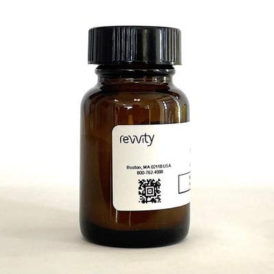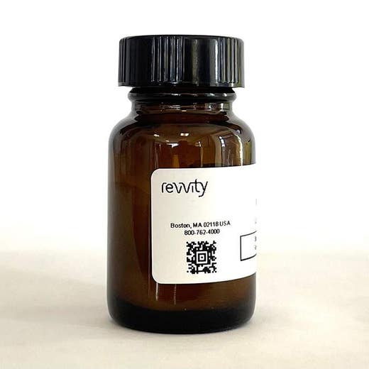
WGA PEI TYPE B PVT SPA Beads, 25 X 2 g

WGA PEI TYPE B PVT SPA Beads, 25 X 2 g


WGA-coated SPA beads, for capturing cell membranes in proximity-based radiometric scintillation assays.
For research use only. Not for use in diagnostic procedures.
| Feature | Specification |
|---|---|
| Bead Type or Material | Polyvinyltoluene (PVT) |
| Surface Treatment |
PEI WGA |
WGA-coated SPA beads, for capturing cell membranes in proximity-based radiometric scintillation assays.
For research use only. Not for use in diagnostic procedures.


WGA PEI TYPE B PVT SPA Beads, 25 X 2 g


WGA PEI TYPE B PVT SPA Beads, 25 X 2 g


Product information
Overview
WGA-coated Polyvinyltoluene (PVT) SPA beads, for capturing cell membranes in proximity-based radiometric scintillation assays. 25 x 2 gram quantity.
The treatment of PVT-WGA SPA beads with positively charged polyethyleneimine (PEI) blocks potential non-specific binding sites on the SPA bead surface. There are two SPA bead types available with PEI treatment. The PVT-WGA-PEI type A SPA beads are treated with PEI prior to the coupling of WGA to the PVT SPA bead. The PVT-WGA PEI type B SPA beads are treated with PEI after the WGA coupling stage. The PVT-WGA-PEI type A and type B SPA beads exhibit different characteristics with regard to the non-specific binding of radiolabelled ligand directly to the SPA bead. Therefore, both bead types should be evaluated when deciding which SPA bead to use and both are included in the Select-a-Bead Kit. The binding capacity of both bead types for cell membrane protein remains 10–30 μg membrane protein per milligram of SPA bead.
SPA Scintillation beads are microspheres containing scintillant which emit light in the blue region of the visible spectrum. As a result, these beads are ideally suited to use with photomultiplier tube (PMT) counters such as the MicroBeta2 or TopCount.
Two types of core SPA Scintillation bead are available - yttrium silicate (YSi) and Polyvinyltoluene (PVT). PVT beads are plastic, larger in size, and stay in suspension longer than the crystalline YSi beads.
Scintillation proximity assay (SPA) is a homogeneous and versatile technology for the rapid and sensitive assay of a wide range of biological processes, including applications using enzyme and receptor targets, radioimmunoassays, and molecular interactions. When 3H, 14C, 33P, and 125I radioisotopes decay, they release β-particles (or Auger electrons, in the case of 125I). The distance these particles travel through an aqueous solution is dependent on the energy of the particle. If a radioactive molecule is held in close enough proximity to a SPA Scintillation Bead or a SPA Imaging Bead, the decay particles stimulate the scintillant within the bead to emit light, which is then detected in a PMT-based scintillation counter or on a CCD-based imager, respectively. However, if the radioactive molecule does not associate with the SPA bead, the decay particles will not have sufficient energy to reach the bead and no light will be emitted. This discrimination of binding by proximity means that no physical separation of bound and free radiochemical is required.
Specifications
| Application |
Drug Discovery & Development
|
|---|---|
| Automation Compatible |
Yes
|
| Bead Type or Material |
Polyvinyltoluene (PVT)
|
| Brand |
SPA Scintillation Beads
|
| Detection Modality |
Radiometric
|
| Format |
Microplates
Tubes
|
| Instrument Compatibility |
Microbeta2 counter
|
| Shipping Conditions |
Shipped Ambient
|
| Surface Treatment |
PEI
WGA
|
| Technology |
Scintilation Proximity Assay
|
| Unit Size |
2 g
|
Resources
Are you looking for resources, click on the resource type to explore further.
Scintillation proximity assay has been successfully applied to receptor binding assays
SDS, COAs, Manuals and more
Are you looking for technical documents related to the product? We have categorized them in dedicated sections below. Explore now.
-
Lot numberANYLot dateSeptember 15, 2023Name


How can we help you?
We are here to answer your questions.


