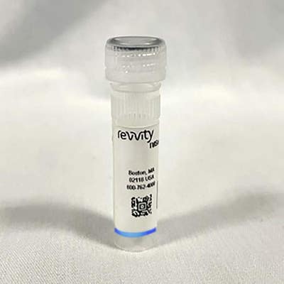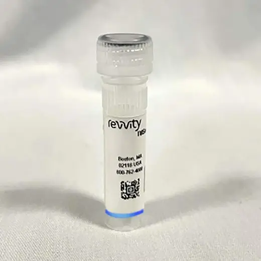
IVISense Hypoxia CA IX 680 Fluorescent Probe (HypoxySense)

IVISense Hypoxia CA IX 680 Fluorescent Probe (HypoxySense)


IVISense™ Hypoxia CA IX 680 (HypoxiSense™ 680) is a carbonic anhydrase IX (CA IX) targeted fluorescent in vivo imaging probe that can be used to image CA IX overexpression in response to hypoxia. Oxygen deprivation is associated with several disease states including angiogenisis, pulmonary disease, inflammation, and cancer.
For research use only. Not for use in diagnostic procedures.
For research use only. Not for use in diagnostic procedures.
| Feature | Specification |
|---|---|
| Wave Length | 680 nm |
IVISense™ Hypoxia CA IX 680 (HypoxiSense™ 680) is a carbonic anhydrase IX (CA IX) targeted fluorescent in vivo imaging probe that can be used to image CA IX overexpression in response to hypoxia. Oxygen deprivation is associated with several disease states including angiogenisis, pulmonary disease, inflammation, and cancer.
For research use only. Not for use in diagnostic procedures.
For research use only. Not for use in diagnostic procedures.


IVISense Hypoxia CA IX 680 Fluorescent Probe (HypoxySense)


IVISense Hypoxia CA IX 680 Fluorescent Probe (HypoxySense)


Product information
Overview
In cancer, hypoxia occurs in tumors as a result of the poor ability of their disorganized vascular networks to deliver blood borne oxygen. IVISense Hypoxia CA IX 680 fluorescent imaging probe detects the tumor cell surface expression of carbonic anhydrase 9 (CA IX) protein, which increases in hypoxic regions within many tumors, especially in cervical, colorectal, non-small cell lung tumors. Pairing IVISense Hypoxia CA IX 680 agent with optical fluorescent imaging technology allows you to image and quantitate tumor sub-regions undergoing hypoxia-related changes, non-invasively and in vivo.
Specifications
| Brand |
IVISense
|
|---|---|
| Fluorescent Agent Type |
Targeted
|
| Imaging Modality |
Fluorescence
|
| Shipping Conditions |
Shipped in Blue Ice
|
| Therapeutic Area |
Angiogenesis
Arthritis
Oncology/Cancer
|
| Unit Size |
1 Vial (10 doses)
|
| Wave Length |
680 nm
|
Resources
Are you looking for resources, click on the resource type to explore further.


How can we help you?
We are here to answer your questions.


