IVISbrite 4T1 Red F-luc-GFP Bioluminescent Tumor Cell line (Bioware Brite)
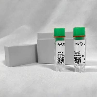
IVISbrite 4T1 Red F-luc-GFP Bioluminescent Tumor Cell line (Bioware Brite)
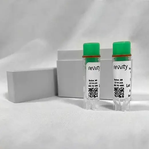


The IVISbrite™ 4T1 Red F-luc-GFP tumor cell line (Bioware® Brite 4T1-Red-FLuc-GFP) is a light-producing and fluorescent cell line derived from 4T1 mouse mammary gland adenocarcinoma. The cells have been stably transduced with the red-shifted firefly luciferase gene from Luciola Italica (Red F-luc) fused to GFP (Green Fluorescent Protein), for a brighter, red-shifted bioluminescent or green fluorescent signal.
For research use only. Not for use in diagnostic procedures.


IVISbrite 4T1 Red F-luc-GFP Bioluminescent Tumor Cell line (Bioware Brite)


IVISbrite 4T1 Red F-luc-GFP Bioluminescent Tumor Cell line (Bioware Brite)


Product information
Overview
IVISbrite 4T1 Red F-luc-GFP Bioluminescent Tumor Cell line
This cell line has very bright bioluminescence, emitting at least 15,000 photons/cell/sec in vitro. The exact number will vary depending on imaging and culturing conditions.
We recommend using this line in Nude and SCID mouse models. An immune response may occur in Balb/c mice.
By emitting intensified, longer wavelength light that is significantly brighter than other firefly luciferases, our bioluminescent oncology cell lines allow you to visualize and monitor the growth of deep tissue tumors in vivo. The GFP fusion also allows for fluorescent imaging in vivo or ex vivo.
After the cells are injected into an appropriate mouse model, the optimized Red F-luc luciferase enables more sensitive in vivo optical detection with less tissue attenuation so you can detect tumor development earlier, and monitor tumor growth and metastases in both subcutaneous and orthotopic models.
- Organism: Mouse
- Tissue: Mammary Gland
- Disease: Adenocarcinoma
- Dual Optical Reporters: Red F-luc and GFP
Specifications
| Brand |
IVISbrite
|
|---|---|
| Cancer Model |
Breast
|
| Imaging Modality |
Bioluminenscence
|
| Luciferase Classification |
Firefly
|
| Shipping Conditions |
Shipped in Dry Ice
|
| Unit Size |
2 vials each containing 1 million cells
|
Resources
Are you looking for resources, click on the resource type to explore further.
SDS, COAs, Manuals and more
Are you looking for technical documents related to the product? We have categorized them in dedicated sections below. Explore now.
-
LanguageEnglishCountryUnited States
-
LanguageEnglishCountryAustralia
-
LanguageEnglishCountryEU
-
Lot numberANYLot dateMay 3, 2024


How can we help you?
We are here to answer your questions.


