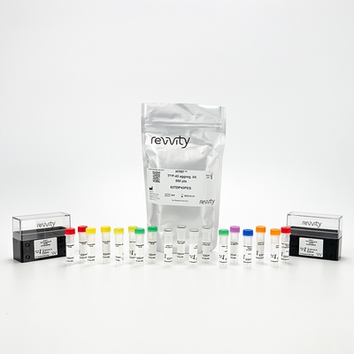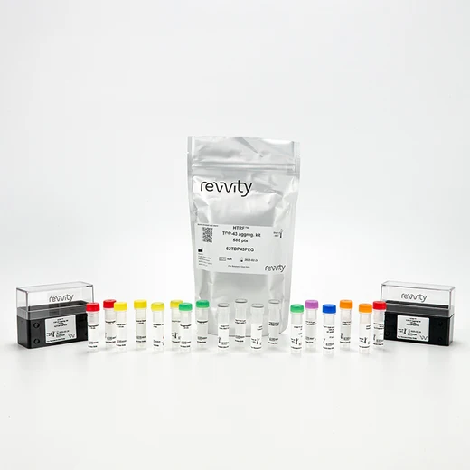

HTRF TDP-43 Aggregation Kit, 500 Assay Points


HTRF TDP-43 Aggregation Kit, 500 Assay Points


This HTRF kit is designed for an accurate measurement of TDP-43 aggregation levels in cellular models.
| Feature | Specification |
|---|---|
| Application | Protein Quantification |
| Sample Volume | 16 µL |
This HTRF kit is designed for an accurate measurement of TDP-43 aggregation levels in cellular models.


HTRF TDP-43 Aggregation Kit, 500 Assay Points


HTRF TDP-43 Aggregation Kit, 500 Assay Points


Product information
Overview
TAR DNA binding protein 43 (TDP-43) is a nucleic acid binding protein involved in RNA-related metabolism. Aggregated TDP-43 has been identified as a hallmark of amyotrophic lateral sclerosis (ALS) and frontotemporal lobar dementia (FTLD), and more widely in several neurodegenerative diseases: TDP-43 proteinopathies. In pathological conditions (mutation or deregulation), TDP-43 forms insoluble inclusion bodies in the cytoplasm of neurons in the brain and spinal cord. The TDP-43 aggregation assay detects human and mouse TDP-43 aggregates rapidly in cellular models.
Specifications
| Application |
Protein Quantification
|
|---|---|
| Brand |
HTRF
|
| Detection Modality |
HTRF
|
| Product Group |
Kit
|
| Sample Volume |
16 µL
|
| Shipping Conditions |
Shipped in Dry Ice
|
| Target Class |
Biomarkers
|
| Technology |
TR-FRET
|
| Therapeutic Area |
Neuroscience
|
| Unit Size |
500 assay points
|
Video gallery

HTRF TDP-43 Aggregation Kit, 500 Assay Points

HTRF TDP-43 Aggregation Kit, 500 Assay Points

How it works
TDP-43 aggregation assay principle
TDP-43 aggregates are measured using a comparison between two HTRF specific signals: the control condition, where soluble TDP-43 only is detected, and the disaggregated condition, where total disaggregated and soluble TDP-43 are assessed. The increase in the disaggregated HTRF signal reflects the level of TDP-43 aggregation.

TDP-43 disaggregation step
Samples containing TDP-43 aggregates require a disaggregation step in order to calculate the level of aggregation of TDP-43 between 2 conditions prior and after sample treatment (sample disaggregation can follow the protocol described on the right). The reagents needed for this step are provided in the kit for maximum convenience. Samples are dispensed directly into the detection (white, low-volume) plate for detection by HTRF® reagents.

TDP-43 aggregation detection
The same control condition sample treated or after the disaggregation step is dispensed directly into the assay plate for detection by HTRF® reagents (384-well low-volume white plate or Revvity low-volume 96-well plate in 20 µl). The antibodies labeled with the HTRF donor and acceptor are pre-mixed and added in a single dispensing step, to further streamline the assay procedure. The assay can be run up to a 1536 well-format by simply resizing each addition volume proportionally.

TDP-43 aggregation assay data analysis
The aggregated ratio of each sample reflects the presence of TDP-43 aggregates in a sample.
In the case of absence of insoluble aggregates, the aggregated ratio is around 1 or below.

Assay validation
Validation of TDP-43 aggregation kit on mouse Neuro2a and human HeLa cell lines
Human HeLa cells were plated at 25,000 and 50,000 cells/well in a 96 well plate, and incubated 24h at 37°C, 5% CO2.
Mouse Neuro2a cells were plated at 50,000 and 100,000 cells/well in a 96 well plate, and incubated 24h at 37°C, 5% CO2.
Cells were either stimulated for 6h with 1 µM of staurosporine (a well established caspase activator cleaving TDP-43 to induce aggregation of C-terminal fragments) for the HeLa cells and 2 µM for the Neuro2a cells, or not stimulated.
After stimulation, the cell culture medium was removed and cells were lysed with 50 µL of supplemented lysis buffer #1 (1X) for 30 min at RT under gentle shaking. Three wells of the same condition were pooled and 2x60 µL of pure lysate for HeLa cells or diluted lysate (at ¼ in supplemented lysis buffer) for Neuro2a cells were transferred into a 96 well plate prior to the disaggregation or control detection step, as follows:
- 10 µL of Disaggregation buffer A or Control detection buffer C added
- Incubation 15 min at RT
- 10 µL of Disaggregation buffer B or Control detection buffer C added
After treatment, 16 µL of each sample (disaggregated or control detection condition) were then transferred into a 384 well low volume white microplate before the addition of 4µL of the HTRF TDP-43 detection reagents. The HTRF signal was recorded after an overnight incubation.
As expected, the staurosporine treatment is associated with an increase in the detection of aggregated TDP-43, whereas in non stimulated samples the ratio is around 1 or below.
Beside demonstrating the compatibility of the assay with human and mouse samples, these results suggest differences in aggregation depending on the cellular background.


Detection of TDP-43 aggregates in overexpressed TDP-43 WT and mutated HeLa cell model
Human HeLa cells (100,000 cells/well) were transfected in a 96 well plate with 200 ng/well of GFP-TDP-43 Wild Type (full length) or GFP-TDP-43 mutated (C Terminal fragment 220-414 aa) using lipofectamine 2000.
After 48h incubation at 37 °C, 5% CO2, cells were stimulated for 6h with 1 µM of staurosporine (a well established caspase activator cleaving TDP-43 to induce aggregation of C-terminal fragments), or not stimulated.
Then the cell culture medium was removed, and cells were lysed with 50 µL of supplemented lysis buffer #1 (1X) for 30 min at RT under gentle shaking.
Three wells of the same condition were pooled and 2x60 µL of diluted lysate (at ¼ in supplemented lysis buffer) were transferred into a 96 well plate prior to the disaggregation or control detection step, as follows:
- 10 µL of Disaggregation buffer A or Control detection buffer C added
- Incubation 15 min at RT
- 10 µL of Disaggregation buffer B or Control detection buffer C added
After treatment, 16 µL of each sample (disaggregated or control detection condition) were then transferred into a 384 well low volume white microplate before the addition of 4 µL of the HTRF TDP-43 detection reagents. The HTRF signal was recorded after an overnight incubation.
As expected, the overexpression of WT or mutated TDP-43 alone increase the aggregated ratio compared to non-transfected cells. The staurosporine treatment triggers an increase in the TDP-43 aggregation ratio, and this effect is stronger on the mutated TDP-43 overexpressed condition.

HTRF TDP-43 aggregation assay compared to Western Blot
Human HeLa cells were grown for 3 days until 80% confluency was reached, in a T175 flask in complete culture medium at 37 °C, 5% CO2. The cells were stimulated with Staurosporine (1 µM) for 6h before lysis with supplemented lysis buffer.
Serial dilutions of the cell lysate were performed using supplemented lysis buffer.
Each dilution was treated in a disaggregation or control condition, as follows:
60 µL of sample,
10 µL of Disaggregation buffer A or Control detection buffer C added,
Incubation 15 min at RT,
10 µL of Disaggregation buffer B or Control detection buffer C added.
16 µL of each dilution (disaggregated or control detection condition) were then transferred into a low volume white microplate before the addition of 4 µL of the HTRF TDP-43 detection reagents. The HTRF signal was recorded after an overnight incubation. Equal amounts of lysates (not disaggregated) were used for a side by side comparison with Western Blot.
As expected, Western Blot shows C terminal fragments (35 kDa) induced by staurosporine treatment which became insoluble and formed aggregates (data not shown). Therefore the HTRF TDP-43 aggregation assay is 2-fold more sensitive than the Western Blot, at least under these experimental conditions.

Simplified pathway
Simplified pathway for TDP-43 aggregation assay
TDP-43 (TAR DNA binding protein 43) is a DNA and RNA-binding protein which plays a crucial role in RNA metabolism. Mainly located in the nucleus, TDP-43 shuttles between the nucleus and the cytoplasm. In pathological conditions, including stress or mutation, there is a mislocalization in the cytoplasm of TDP-43 which can be cleaved in C terminal fragments, thus leading to an accumulation of ubiquitinated and hyperphosphorylated toxic aggregates and then insoluble inclusion bodies. Mislocalized soluble TDP-43 becomes ubiquitined to be degraded by the proteasome, whereas autophagy tries to remove insoluble aggregates when it is still possible.
The involvement of TDP-43 aggregates at the cellular level has been studied in numerous mechanisms maintaining RNA metabolism and cell homeostasis, such as proteasomal activity, autophagy, or mitotoxicity…
The presence of TDP-43 inclusion bodies in neurodegenerative diseases, such as Amyotrophic lateral sclerosis (ALS), FrontoTemporal Dementia (FTD), or Alzheimer’s Disease, suggests an important role of TDP-43 in neurodegeneration processes and opens new avenues for therapeutic discoveries.

Resources
Are you looking for resources, click on the resource type to explore further.
Emerging pathways to neuroinflammation and neurodegeneration
Neurodegenerative diseases, such as amyotrophic lateral sclerosis...
Discover the versatility and precision of Homogeneous Time-Resolved Fluorescence (HTRF) technology. Our HTRF portfolio offers a...
This guide provides you an overview of HTRF applications in several therapeutic areas.
Discover the power of HTRF for TDP-43 phosphorylation and aggregation investigations
The trans-activator response (TAR) DNA-binding...
Targeted protein degradation (TPD) offers a promising approach for treating neurodegenerative diseases by selectively degrading...
Loading...


How can we help you?
We are here to answer your questions.


